Soft Imaging System의 디지털 카메라는 디지털 이미지 획득의 기대에 부응하여 꾸준히 대응하고 있습니다.
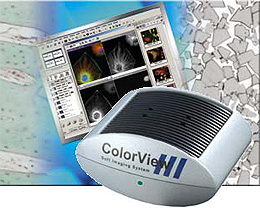
ColorView III는 광학 현미경을 위한 칼라 카메라 시리즈의 최신작 입니다.
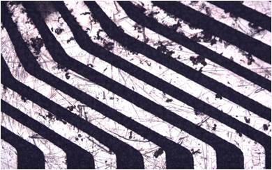
ColorView III는 생명과학(Life science) 및 Biomedicine의 모든 응용 분야 뿐만 아니라 재료 과학 (Materials sciences) 분야에 동등하게 적용할 수 있습니다.
Extraordinary features…
are what make the 5 MegaPixel color camera ColorViewIII so distinctive. Alongside its extremely high resolution, the Peltier-cooled camera has a high dynamic range, high frame rates, partial read-out and color binning. The color fidelity and superior contrast of acquisitions made using the ColorViewIIIare what users find so compelling. At the same time, the camera has an excellent signal-to-noise ratio. The standard C-mount adaptor ensures that installation is easy on all common microscopes. Data transmission to the PC is via FireWire™ interface (IEEE 1394).
Perfect images…
easy to use and full software operation due to integration of all camera functions within the analySIS® image-analytical software. “Digital Live Processing” is integrated into analySIS® and offers numerous real-time functions which ensure that the camera’s entire dynamic range is exploited. Furthermore, analySIS® has IntX (Intelligent eXposure) – an integrated solution for easy-to-use and automatic exposure control. This makes acquiring top-quality images at the mere click of a button easy for all users. No extensive technical background is required.
And after acquisition…
the ColorViewIII’s full integration into the analySIS®software provides all the capabilities and advantages of advanced image processing. There’s an extensive image-labeling-and-filtering library, professional particle analysis, well-structured image and data archiving as well as report generation for professional documentation.
Slides by Volker Neureuther, Ingolstadt, Germany
Depending on options, analySIS® expansion level and
CC-12는 어떠한 광학 현미경 어플리케이션에도 대응하는 고해상도 12비트 칼라 CCD 카메라 입니다.
FireWire 기술 (IEEE1394)에 의한 Peltier 냉각 및 전력을 공급받으며 어떠한 저조도 환경에서도 최적의 신호대 잡음비를 제공해 줍니다.
CC-12는 “analySIS Five” Image Analyzer Software에 완전히 통합됩니다.
Digital solutions – Developed to be exceptional
The series of light microscope cameras by Soft Imaging System has been designed to meet the highest digital-imaging acquisition demands for all areas of the microscopy field.
The CC-12 is the successor to the ColorView12. This 12-bit, Peltier-cooled and FireWire™(IEEE 1394) equipped color digital camera is the ideal introduction to digital image acquisition in the field of light microscopy.
All functions of the camera can be completely controlled and operated via the analySIS®image-analytical software. No matter what current acquisition conditions are, real-time functions guarantee that the entire dynamic range is taken advantage of. This ensures the user optimal contrast all the time.
Following acquisition, CC-12’s total integration into analySIS® provides the full range of options and advantages of the latest in image processing and analysis, from image labeling to archiving, report generation and e-mailing all the way on to photo-realistic printouts – and there’s no more need for the darkroom.
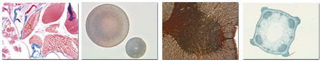
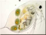
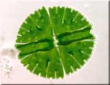
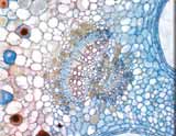
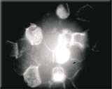
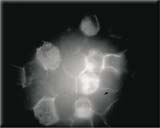
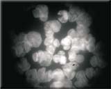
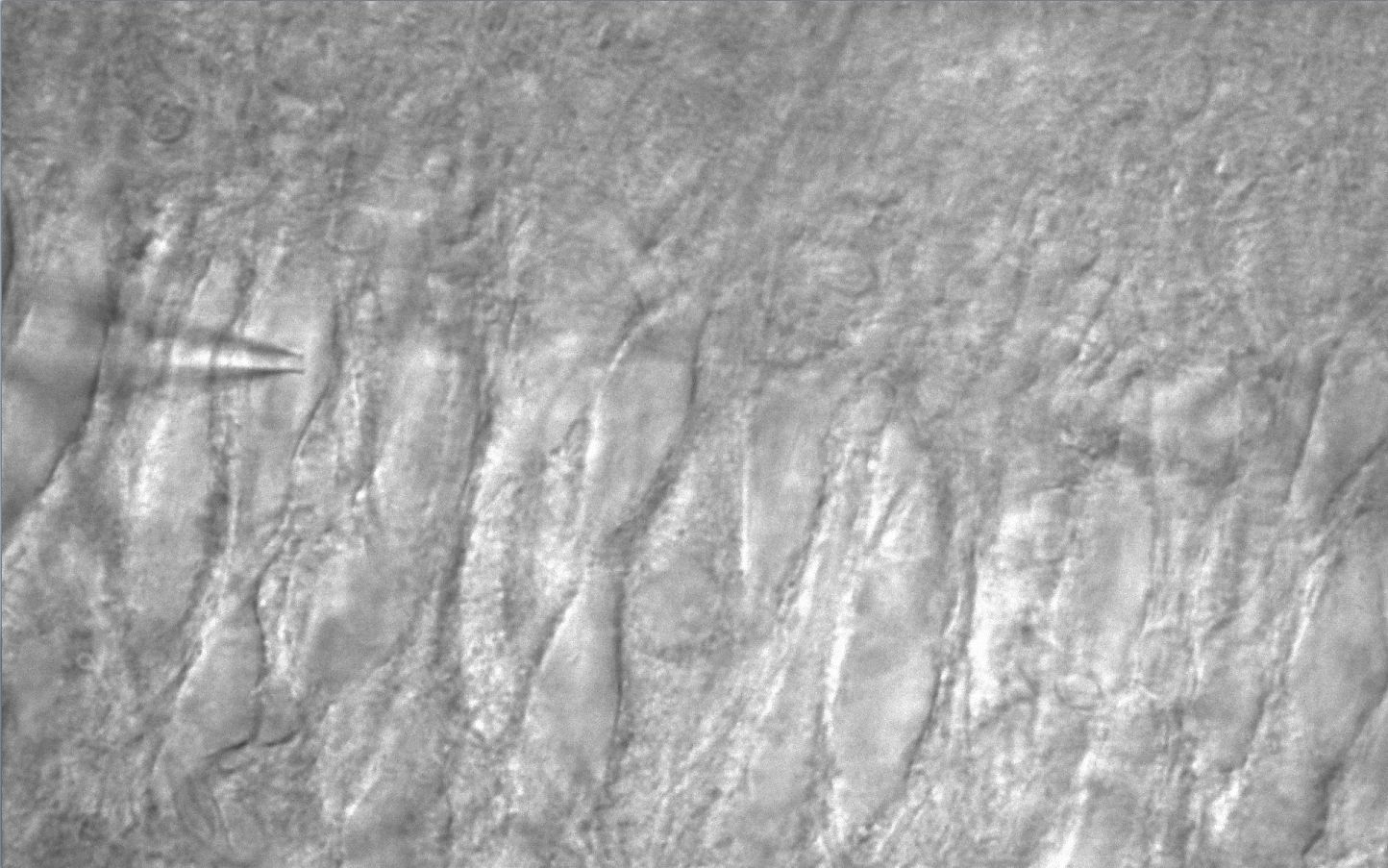
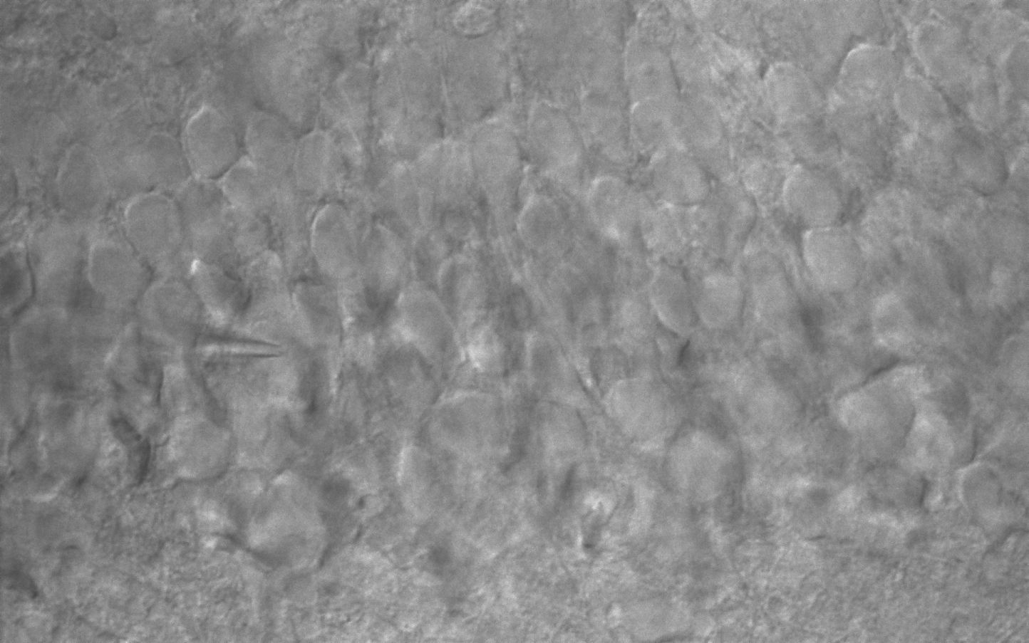
CC-12 mounted onto Olympus BX60F daphnia longispina (long-spined water flea) with eggsCC-12 mounted onto Olympus BX60F micrasterias rotata (a desmid – single-celled green algae)CC-12 mounted onto Olympus BX60F stem axis of tulipa gesneriana (garden tulip)
High Resolution
The CC-12 camera has a resolution of 1300 x 1030 pixels. This is three times greater than that of a regular video camera.
A camera that’s low-noise and cooled
Highly efficient readout technology (Correlated Double Sampling) coupled with Peltier-cooling of the CCD chip results in images of superior signal-to-noise ratio.
Variable exposure times
Highly sensitive CCD elements are even capable of detecting signals that are extremely weak. The electronic shutter offers variable exposure times ranging from 100 µs to 160 seconds.
High frame rate
The high speed ADC (Analog-Digital Converter) working at a clock rate of 20 MHz in full 12-bit dynamic range is able to perform double sampling even at a readout rate of 20 MHz. Various frame rates are supported by this camera. For example, the camera can be set to acquire at a high frame rate of more than 22 fps at TV resolution using 2x binning. View your zoomed-in sample, locate the area of interest and focus – all conveniently onscreen. No longer are you forced to trade off speed for quality. For acquisition the system switches automatically into the high-resolution mode. This avoids bleaching of your fluorescence specimen and offers optimal performance when setting parameters.
FireWire™ Interface (IEEE 1394)
FireWire™ technology guarantees that the CC-12 installation is easy on any PC or laptop equipped with a FireWire™ port. The days when you were limited to a frame grabber and just a single camera are history. FireWire™ technology enables you to use multiple cameras on the same PC.
Peltier-Cooling
Image noise is generally the result of one of two things: either the CCD chip is not being cooled to a low enough temperature (known as ”dark current”), or due to mechanical vibrations. For the CC-12, thermal noise and instability is not a problem because the CCD chip is Peltier-cooled and stabilized at 10°C, resulting in a very high signal-to-noise ratio. Noise is further suppressed by the application of a highly efficient digital readout technique known as Correlated Double Sampling.
Installation is easy
The CC-12 can be mounted onto all light microscopes with a C-mount adaptor. Plus, you only need one cable for getting data and power to the PC’s FireWire™ port. No more clutter and no ’octopus’ of cables getting in your way.
Real-time functions CC-12 is fast. Put this in combination with the high speed of today’s CPU’s, and you’ve got an attractively broad range of real-time functions within analySIS® available to you. These include automatic contrast control, automatic white balance and histogram display.
Analyzing images
A vast library of text, graphic and editing functions is available for labeling images. Special filters and professional particle analysis assist in more extensive investigation of images. All this makes it simple to obtain reliable and reproducible results quickly.
No more darkroom
Photo-quality printouts can be obtained following acquisition without any need for a darkroom nor its developing chemicals. And these photo-quality printouts are in your hands in minutes.
Archiving analySIS® offers you a powerful, fully network-able, image-archiving system that handles all the images and data generated during the process of image acquisition and documentation. Images can be stored along with text, sheets and other graphics as records for complete documentation of tasks and processing steps. Input, display and query masks can be defined independently.
Automatic report generation
Now you can produce multi-page reports quickly and efficiently. Select multiple images in the image database and insert them all into the report via a single command. In addition to the images themselves, you can have information from any database field automatically included in reports. Automatic scaling, detail zooms, and more – all available to optimize the way you work with images. All documents generated using the analySIS® software can be inserted into reports. Use the report generator to print out images, related measurement sheets, and diagrams – all on the same page. This report generator provides you with the utmost flexibility for page layout and design. You set up your own templates exactly the way you want them to be. Templates need to be created just once. Templates are what your reports are based on and ensure that the appearance of your documents is uniform. Use the RTF Export function to export your reports to MS Word for continued editing.
CC-12 and mia
Automatic pattern recognition means you can use mia to montage multiple component imagesacquired with the CC-12into a single, high-resolution image.
This is tremendously useful, eg, when you wish to display a sample in its entirety at high resolution but the microscope is only capable of showing a portion of the sample at such high resolution. All you need to do is define image size and resolution. The rest is taken care of automatically.
Software control of all camera functions
Manufacturer programmable RISC CPU + FPGA via firmware download
Multi-threading code support on multi-CPU PC
Depending on options, analySIS® expansion level and
Real-time automatic contrast control
Real-time automatic white balance
Black balance
Sharpness monitor
Specifications
Image Device
2/3 inch Color CCD Sensor (Effective area 8.9 x 6.7 mm array)
이 제품은 극도로 높은 감도, 저 노이즈, 고해상도의 정량화할 수 있는 이미지를 요구하는 응용분야에 이상적입니다.
F-View II 는 FireWire 기술 (IEEE1394)에 의한 Peltier 냉각 및 전력을 공급받으며 FISH 혹은 Single band-pass 필터를 사용하는 다중 노출 형광 응용분야를 위해 특별히 설계되었습니다.
F-View II 는 “analySIS Five” Image Analyzer Software에 완전히 통합됩니다.
Digital solutions – Developed to be exceptional!
Soft Imaging System’s digital cameras are designed to meet the steadily rising demands on digital image acquisition for all areas of microscopy. And so is the newly developed 12-bit, cooled CCD color camera F-ViewII, our latest 12-bit, Peltier-cooled, FireWire™-driven solution for fluorescence microscopy.
The search mode enables you to rapidly locate and conveniently focus in on the area of your sample of interest. This prevents any unnecessary bleaching of the sample.
The camera will automatically switch over to the high-resolution mode for acquisition. F-ViewII can also be easily operated using a laptop via the FireWire™ port.
All camera functions are fully controlled by the analySIS® image-processing software. Real-time functions enable the use of the entire dynamic range under all conditions and guarantee the best contrast.
F-ViewII’s integration into analySIS® provides all the capabilities and advantages of state-of-the-art digital image processing and analysis, ranging from image labeling, archiving, report generation and e-mailing, as well as photo-realistic printouts without the darkroom.
High Resolution
The CCD chip of the F-ViewII has 1376 x 1032 pixel resolution. The 12-bit dynamic range means that even acquisition of images with very bright and faint regions is dynamically optimal.
High sensitivity
CCD elements are so sensitive that they have no trouble detecting even the weakest of signals. The electronic shutter offers variable exposure times ranging from 100 µs to 160 seconds.
High frame rate
The high speed ADC (Analog-Digital Converter) working at a clock rate of 20 MHz in full 12-bit dynamic range is able to perform double sampling even at a readout rate of 20 MHz. Various frame rates are supported by this camera. For example, the camera can be set to acquire at a high frame rate of more than 22 fps at TV resolution using 2x binning. View your zoomed-in sample, locate the area of interest and focus – all conveniently onscreen. No longer are you forced to trade off speed for quality. For acquisition the system switches automatically into the high-resolution mode. This avoids bleaching of your fluorescence specimen and offers optimal performance when setting parameters.
FireWire™ Interface (IEEE 1394)
FireWire™ technology guarantees that the F-ViewII installation is easy on any PC or laptop equipped with a FireWire™ port. The days when you were limited to a frame grabber and just a single camera are history. FireWire™ technology enables you to use multiple cameras on the same PC.
Peltier-Cooling
Image noise is generally the result of the CCD chip not being cooled to a low enough temperature (“dark current”). For the F-ViewII, thermal noise is not a problem because the CCD chip is Peltier-cooled and stabilized at 10°C. Noise is further suppressed by the application of a highly efficient digital readout technique known as Correlated Double Sampling (CDS).
Compact design
With its elegantly compact, newly designed housing, the F-ViewII can be mounted onto all light microscopes with a C-mount adaptor. No other interfaces or adaptors are necessary. Plus, you only need one cable for getting data and power to the PC’s FireWire™ port. No more clutter and no “octopus” of cables getting in your way.
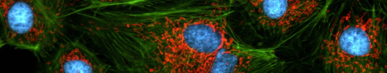
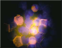
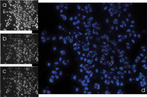
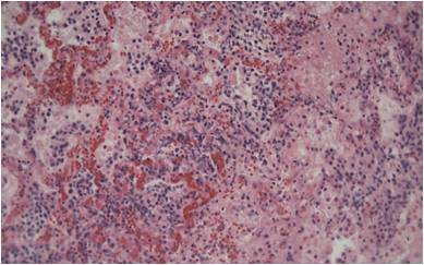
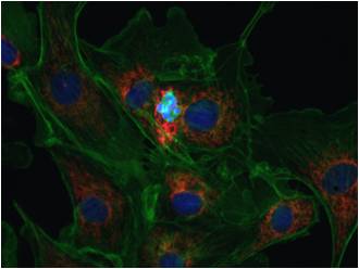
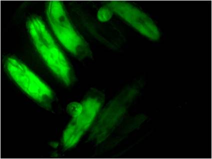
DAPI, PE, APC, PerCP & FITC image acquisitions of stem cells: combined acquisitions done via themFipanalySIS® add-in
source: Dr. Hans Wessels
University Clinic for Children, Tübingen, Germany
Real-time functions F-ViewII is fast. Put this in combination with the high speed of today’s CPU’s, and you’ve got an attractively broad range of real-time functions within analySIS®available to you. These include automatic contrast control, sharpness monitoring and histogram display.
Analyzing images
A vast library of text, graphic and editing functions is available for labeling images. Special filters and professional particle analysis assist in more extensive investigation of images. All this makes it simple to obtain reliable and reproducible results quickly.
No more darkroom
Photo-quality printouts can be obtained following acquisition without any need for a darkroom nor its developing chemicals. And these photo-quality printouts are in your hands in minutes.
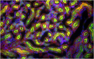
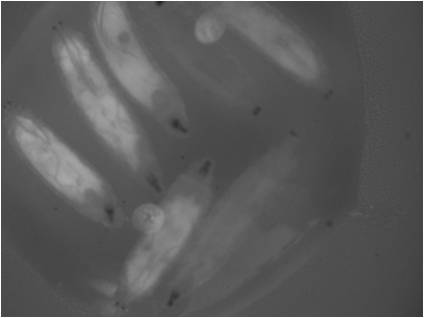
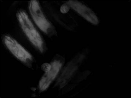
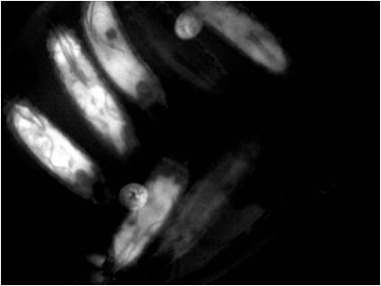
a) Her2 gene (Spectrum Orange)
b) DNA (DAPI)
c) Chromosome (Spectrum Green)
d) Combination with mFip
Specimen: Norbert Wey
University of Zürich
Department of Pathology
Switzerland
Archiving analySIS® offers you a powerful, fully networkable, image-archiving system that handles all the images and data generated during the process of image acquisition and documentation. Images can be stored along with text, sheets and other graphics as records for complete documentation of tasks and processing steps. Input, display and query masks can be defined independently.
Automatic report generation
Now you can produce multi-page reports quickly and efficiently. Select multiple images in the image database and insert them all into the report via a single command. In addition to the images themselves, you can have information from any database field automatically included in reports. Automatic scaling, detail zooms, and more – all available to optimize the way you work with images.
All documents generated using the analySIS® software can be inserted into reports. Use the report generator to print out images, related measurement sheets, and diagrams – all on the same page. This report generator provides you with the utmost flexibility for page layout and design. You set up your own templates exactly the way you want them to be. Templates need to be created just once. Templates are what your reports are based on and ensure that the appearance of your documents is uniform. Use the RTF Export function to export your reports to MS Word for continued editing.
Software control of all camera functions
Manufacturer programmable RISC CPU + FPGA via firmware download
Multi-threading code support on multi-CPU PC
Depending on options, analySIS® expansion level and
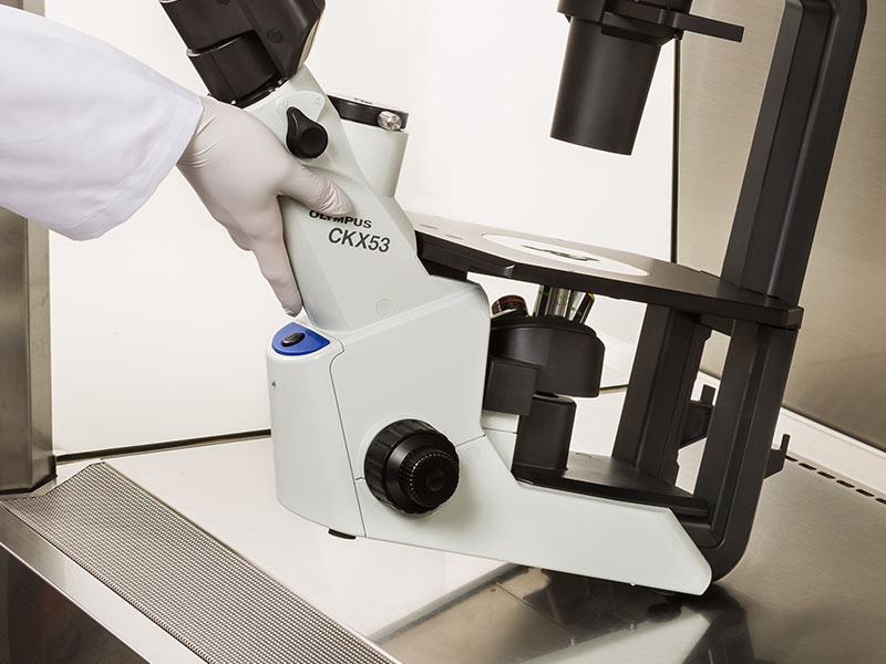
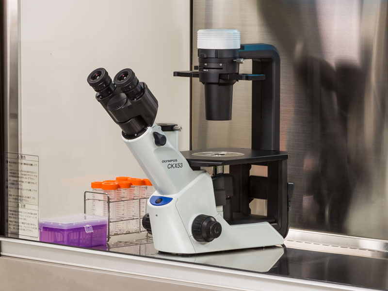
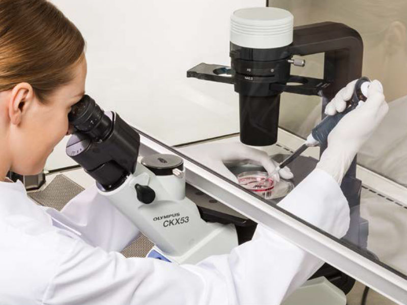
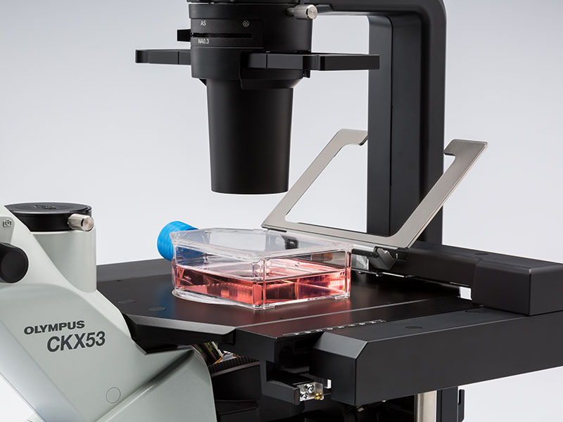
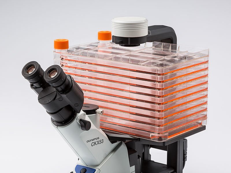
향상된 이미지 품질과 인체공학 설계로, Olympus CKX53은 라이브 셀 관찰, 세포 샘플링 및 처리, 이미지 캡처, 그리고 형광 관찰을 포함한 다양한 세포 배양 샘플에 뛰어난 성능과 효율적인 관찰 흐름을 제공합니다.
라이브 셀 관찰
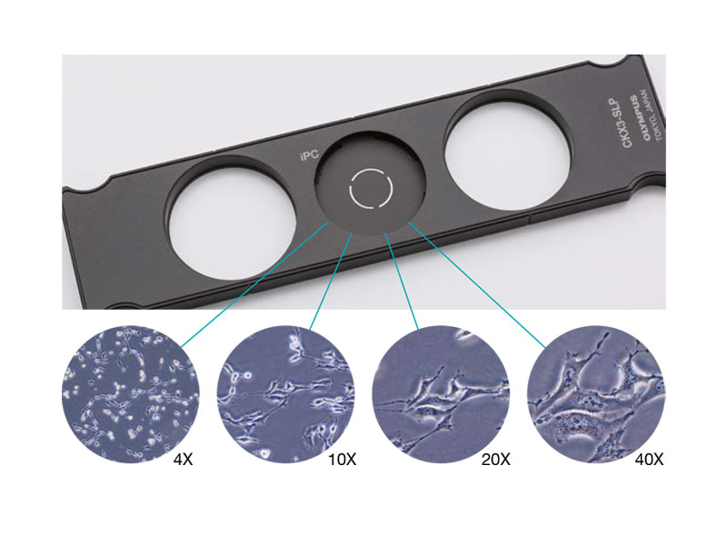
통합 위상차( iPC : Integrated Phase Contrast ) 현미경
CKX53 iPC 시스템을 이용하여 대물렌즈 배율( 4x, 10x, 20x , 40x ) 변경시, 콘덴서 측의 위상차 링슬릿의 연동 변경이 필요 없게 되어 효율적인 관찰 작업이 가능하고, 또한 위상차 설정이 틀어짐에 대한 수시 조정이 필요없어 언제나 선명한 샘플 관찰이 가능합니다.
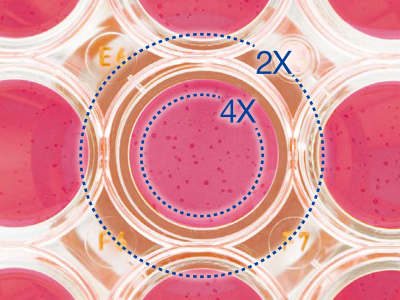
2X 배율, FN 22의 대물렌즈로 선명하고 넓은 시야
PLN2X 대물렌즈를 위한 링 슬릿, CKX3-SLPAS는 직경 11 ㎜ , 시야수 22 ㎜ 를 갖습니다.
2X 대물렌즈는 다른 대물렌즈 보다 확연히 높은 contrast를 제공하여, 투명한 샘플도 명확하게 식별할 수 있습니다. 예를 들어, 96-웰 마이크로 플레이트 관찰시, 넓은 시야로 인하여 스테이지를 움직이지 않고 웰의 모든 세포를 관찰 할 수 있습니다.
IVC (Inversion Contrast) 기술을 사용한 3D 셀 관찰
새로 개발된 IVC 기술로, 위상차보다 시야 심도는 좁아지며, 개체의 모양이나 투명도와 관계없이 삼차원 이미지를 선명하게 합니다. 또한, IVC 관찰은 후광 효과나, 방향성 있는 그림자를 배제하여, 개체의 선명한 관찰을 가능하게 합니다. * 10X 대물렌즈 (PLCN10X, CACHN10XIPC)는 새로운 IVC 관찰에 사용할 수 있습니다.
Glass Heater for microscope
TPi-CKX53X ( Thermo Glass Plate )
Microscope:Olympus CKX53 series
Applicable stage: XY mechanical stage CKX3-MVR
Setting range: ambient ~ 60℃
Plate dimension: W190 x D138㎜
Heating area: W174 × D127㎜
Glass thickness: 0.5 ㎜
형광 관찰 (Fluorescence Microscopy)
다양한 형광 시약과 선명한 시야
100 W 수은 램프 (U-LH100HG), 130 W 고압 수은 램프 (U-HGLGPS), 그리고 타사(3rd Party) LEDs*와 같은 여러 통합 광원을 이용하여 형광 이미지를 선명하게 관찰할 수 있습니다. 일반 연구용 형광현미경 IX3 및 BX3 에서 사용하는 미러 유닛을 동일하게 사용 할 수 있습니다.
3개의 형광미러 유닛을 장착할 수 있으나, Bright Field 관찰과 위상차 관찰에 영향을 줄 수 있으니 유닛 선택시 고려할 필요가 있습니다.
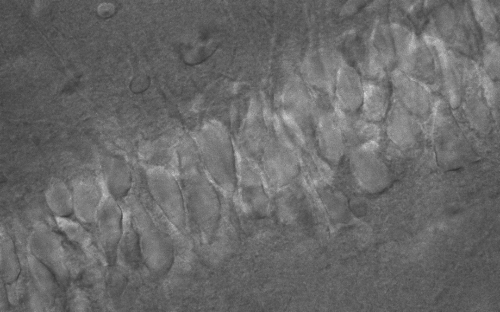
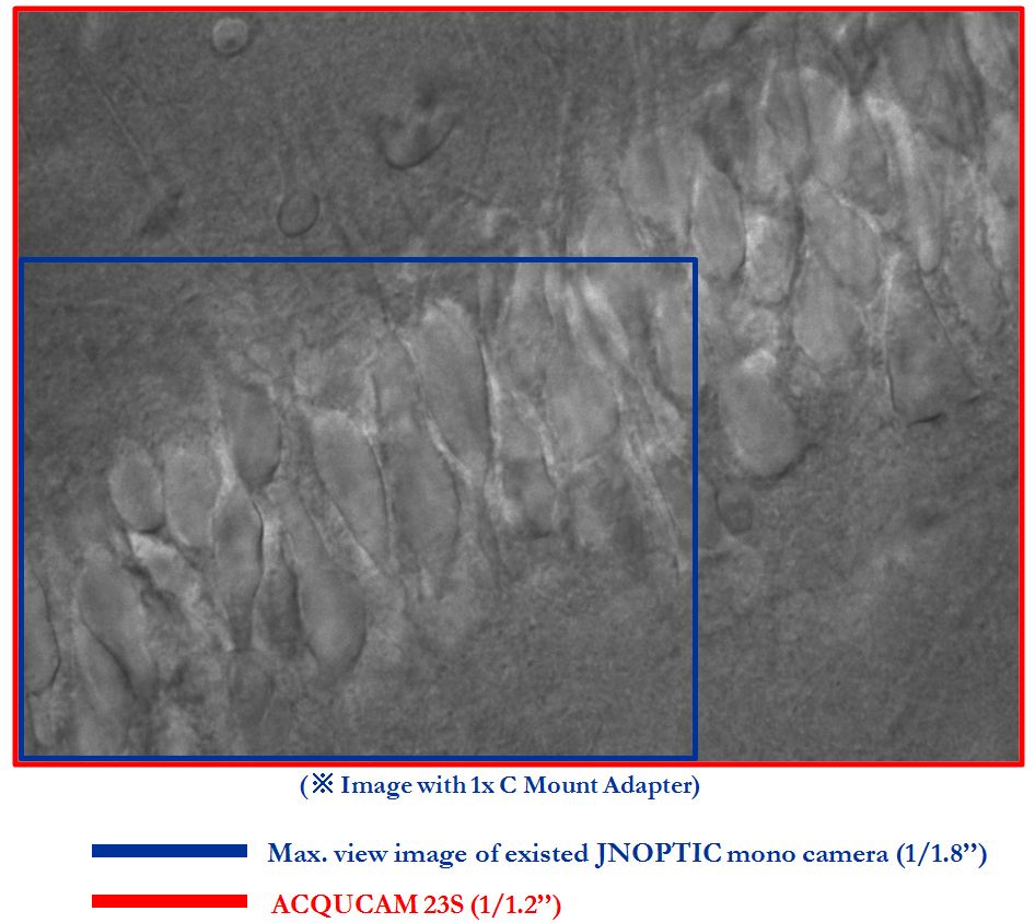
Image taken by AcquCAM 23GR2 with LUCPlanFLN40x Ph2, 1x Adapter, CKX53
밝은 조건에서 높은 Contrast
“Umbra Shield”는 특히 CKX53을 사용한 형광 관찰을 위해 설계되었습니다. 차단막은 실내 광원을 효과적으로 차단하여 형광의 대비를 향상하여 밝은 실험실 조건에서도 선명한 형광 관찰이 가능합니다. 위상차를 사용하는 경우, Umbra 차단막을 들어 올려 표본에 빛을 통과시킬 수 있습니다.
CKX53 현미경은 UV 차단 코팅 덕분에 UV 살균 공정 중에 그대로 둘 수 있습니다. 이 시스템은 약 7kg (15.4lb)으로 이전 모델보다 가볍고 설치 공간이 더 작기 때문에 실험실 공간을 덜 차지합니다. 또한, 한 손으로 현미경을 움직일 수 있으며 관찰 경통의 목 부분을 이용하여 쉽게 운반 할 수 있습니다.
멸균 벤치 환경에서 간편한 세포 샘플링
CKX53의 접안렌즈와 광축/포커스 노브 사이의 거리가 짧으므로 작업자의 손의 위치를 자연스럽게 잡을 수 있어서 초점 및 셀 샘플링이 용이합니다.
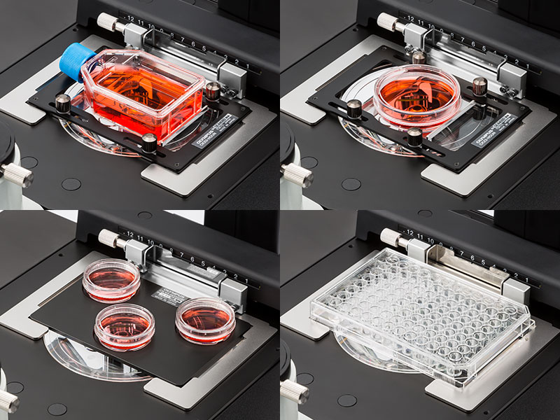
다양한 세포 배양 용기를 사용할 수 있습니다.
CKX53의 공용 홀더로, 디쉬, 마이크로플레이트, 플라스크를 포함한 다양한 용기에서 배양된 세포를 확인하기 쉽습니다. 옵션 홀더가 부착되면, 최대 세 개의 35㎜ 배양 용기를 스테이지에 장착할 수 있습니다. 또한, 다양한 마이크로플레이트를 별도의 홀더 없이 다룰 수 있습니다.
다층 조직 플라스크(Multi-Layer Tissue Flask)를 위한 종합적인 관찰
CKX53의 폭과 탈착 가능한 콘덴서로 다층 조직 플라스크와 같은, 최대 190 ㎜ 높이의 배양 용기도 볼 수 있습니다. PLCN4X 대물렌즈의 우수한 초점 심도로 다층 조직 플라스크 내 바닥 두 개의 층의 세포를 빠르고 편하게 관찰할 수 있습니다.
다양한 용기를 사용하여 관찰 유연성 증대
홀더 암을 들어 올려서 수동으로 세포 배양 용기를 배치할 수 있습니다. 또한, 스테이지는 좌우로 최대 70 ㎜ 까지 확장할 수 있습니다.
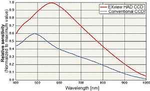
The displays live digital images with gradual smoothness and combines exceptional resolution with faithful color reproduction.The new sensors feature a global shutter function and able to capture a high-speed moving image without focal plane distortion. High-speed processing, low noise and low power dissipation by using column-parallel A/D conversion. equipped with trigger mode, and the external pulse can control accumulation time. The Sensor also have a pulse output function to indicate respective conditions during shutter operation and can be coordinated with peripheral circuits. High-definition images can be displayed live at a rate of high frames per second. without compression. Such imaging quality enables even the finest cellular regions to be observed clearly and distinctly without deterioration. While focusing is made stress free.
Universal camera- wide range of image acquisition is available
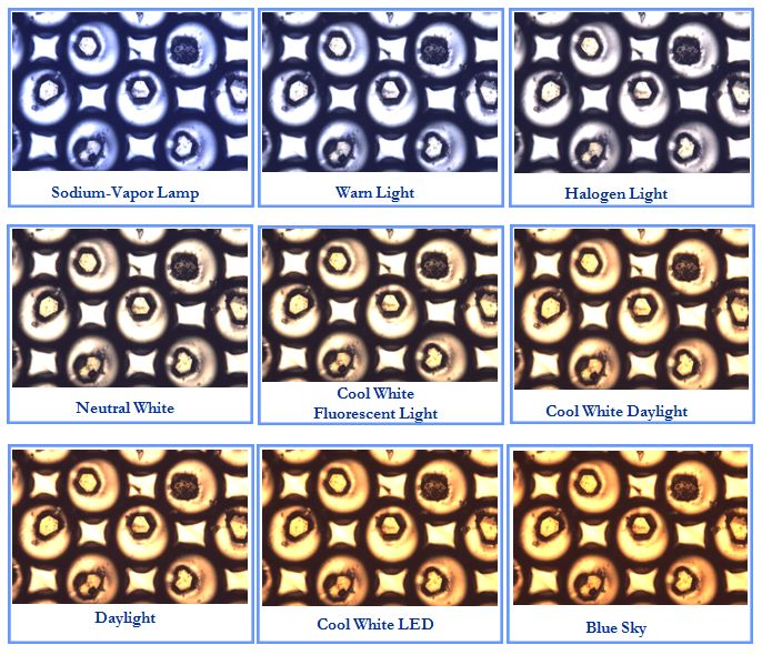
Setting function of variety color images
With wide range of color temperature setting function, you can obtain a clear image of high sensitivity in accordance with the characteristics of samples you want to observe.
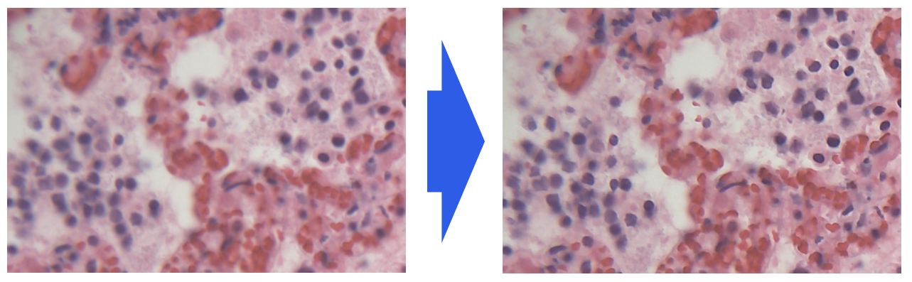
Live sharpness adjustment
You can observe and obtain sharp images by applying the sharpness function even when the live video output
AcquCAM Silver3 camera has 1/1.8 CCD Color Sensor with USB 3.0 and works with resolution of 1600×1200 @ 20fps. Self-developed WDM-based drivers provide stable stream and C & CS mount lens can used in.
2 Mega pixel CCD USB 3.0 camera
High sensitive monochrome
Preview software provided (JNOPTIC Capture)
Software compatible with Windows XP/ Vista/ 7/ 8 operating systems
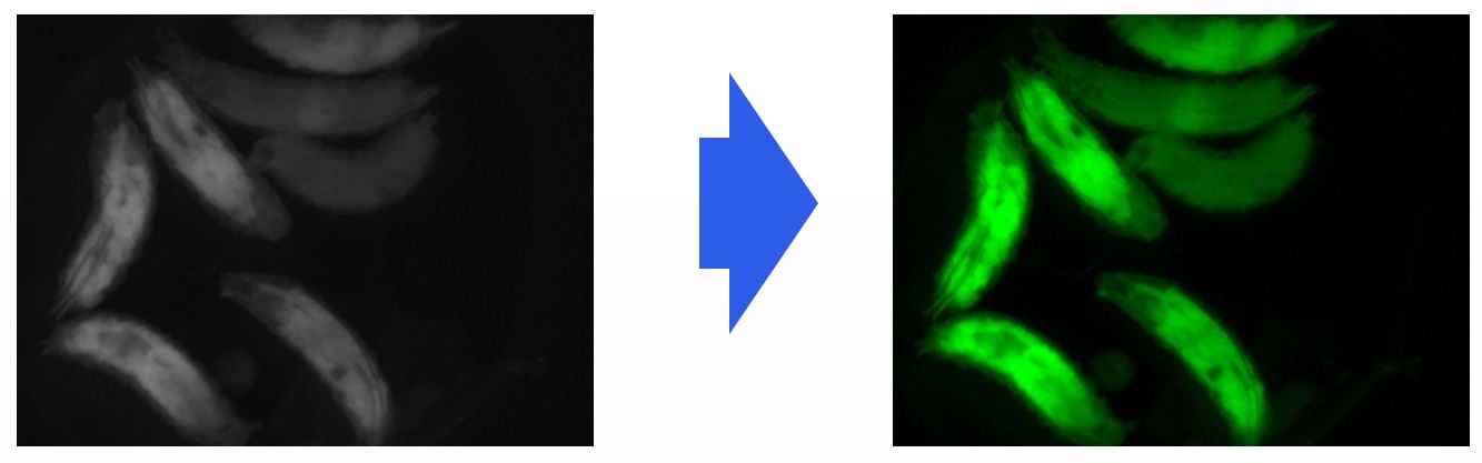
Apply Pseudo color for live fluorescence
Increased resolution by applying PSEUDO COLOR function when observing fluorescence image.
Apply Pseudo color for live fluorescence
Shooting images of fluorescence sample
Emphasis function on variety of images
1. Original image
2. After application of BLACK BALANCE
3. After application of AUTO LEVEL
4. After application of PSEUDO COLOR
Getting fast and clear image with general light source
Shooting by stereo microscope and LED lighting source