The brand new Kinetix family of back-illuminated sCMOS cameras delivers a framerate and field of view unmatched by any other sCMOS camera. KINETIX CAMERA
The small, 4.25 µm pixels provide highly detailed images across the imaging plane, which allows for the highest resolution when using lower magnification objectives.
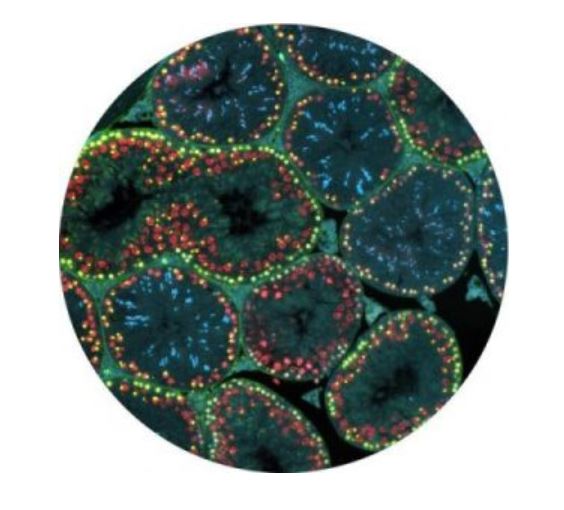
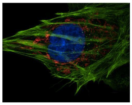
sCMOS Sensitivity
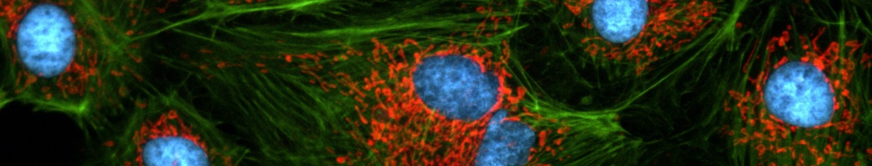
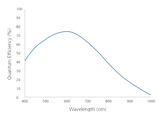
The low noise and high quantum efficiency across a wide range of wavelengths produces higher quality images with lower exposure times than conventional CCD cameras.
Advanced Triggering
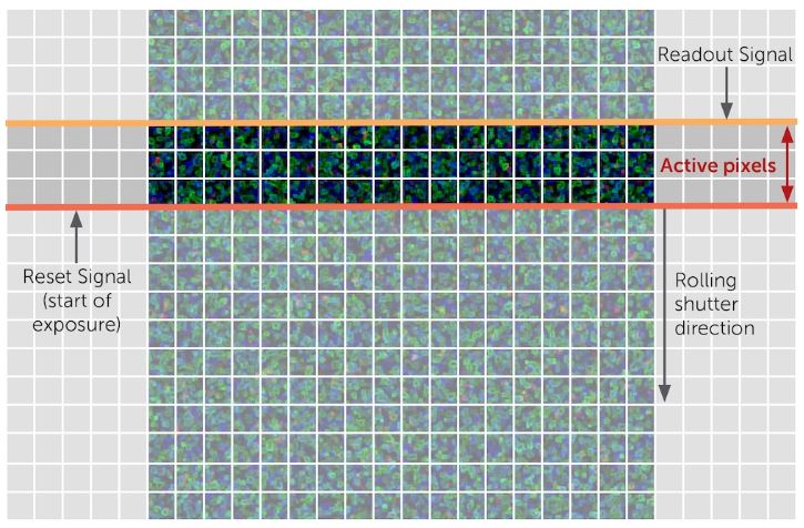
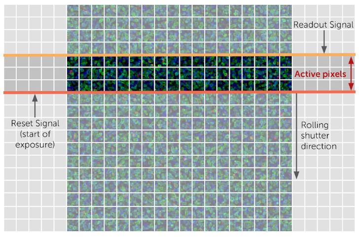
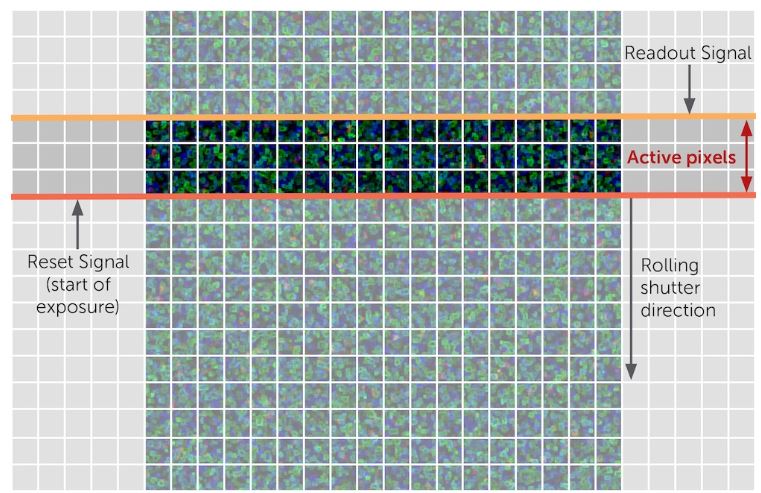
Programmable Scan Mode provides increased control over the rolling shutter exposure and read-out functionality of CMOS sensors by providing access to the sensor timing settings to allow optimization around applications that require control over the line time.
Superior Background Quality
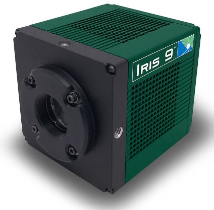
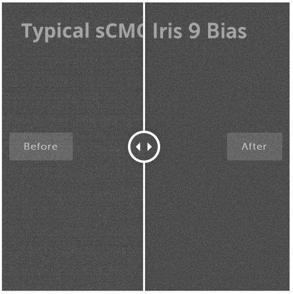
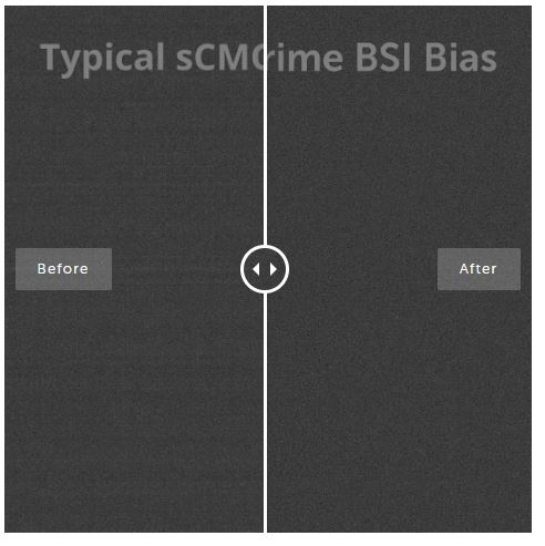
The Iris 9 features Pattern Noise Reduction Technology and Correlated Noise Reduction Technology to ensure that it delivers clean, pattern-free images with minimal pixel defects, delivering improved image quality in low light conditions.
Compact Form Factor
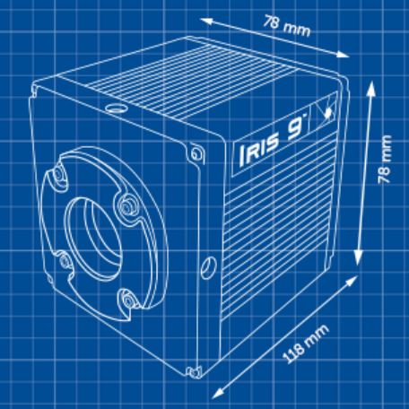
The Iris 9 is a compact 76 x 76 x 88 mm with optimized cooling for the size, ideal for integration into new or existing configurations.
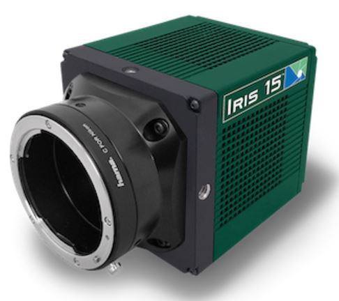
The larger format 25 mm sensor of the Iris 15 is designed to increase throughput, maximize the amount of data captured and take full advantage of new, larger field of view microscopes.
High Resolution
The small, 4.25 µm pixels provide highly detailed images across the imaging plane, which allows for the highest resolution when using lower magnification objectives.
Advanced Triggering
Programmable Scan Mode provides increased control over the rolling shutter exposure and read-out functionality of CMOS sensors by providing access to the sensor timing settings to allow optimization around applications that require control over the line time.
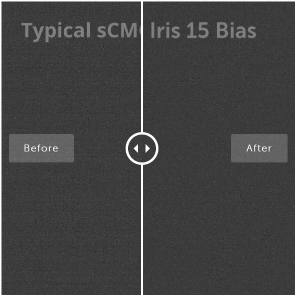
Superior Background Quality
The Iris 15 features Pattern Noise Reduction Technology and Correlated Noise Reduction Technology to ensure that it delivers clean, pattern-free images with minimal pixel defects, delivering improved image quality in low light conditions.
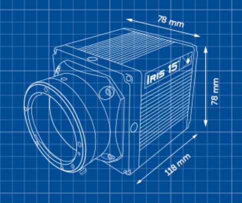
Compact Form Factor
The Iris 15 is a compact 78 x 78 x 108 mm with optimized cooling for the size, ideal for integration into new or existing configurations.
UV(200-400nm), Visible(400-700nm), Near Infrared(700-1000nm)
Read Noise(median)
1.2 e-
Sensor Area
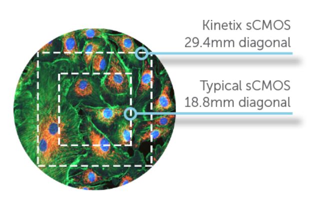
20.8mm X 20.8mm , 29.4mm Diagonal
Frame Rate
400fps @ 8bit / 90fps @12bit / 83fps @ 16bit
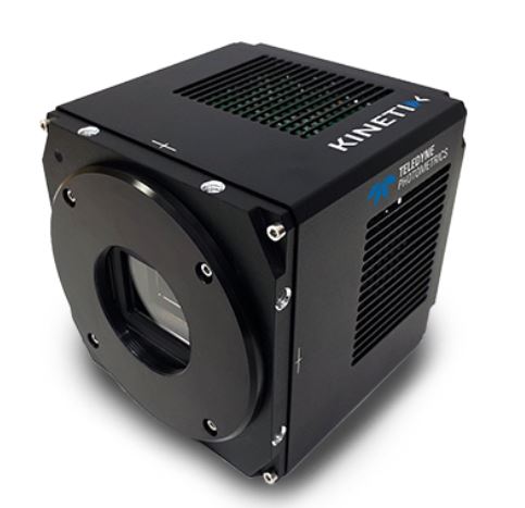
Kinetix
10.2M pixels (3200×3200)
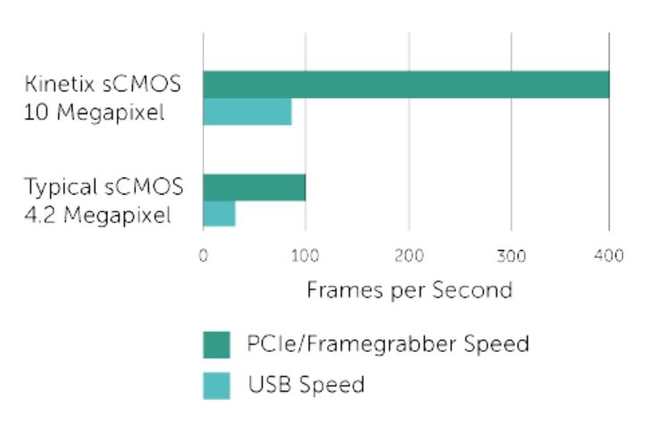
Extreme Speed
Taking advantage of an 8-bit readout mode, the Kinetix sCMOS delivers a tremendous 400 frames per second (fps), full frame with a 29.4 mm diagonal field of view. The optimized line time allows the speed to significantly outperform typical sCMOS devices, delivering over 4000 megapixels/second – an almost 10-fold improvement
Large Field Of View
The 29.4 mm square sensor of the Kinetix is designed to increase throughput, maximize the amount of data captured in a single frame and take full advantage of new, larger field of view microscopes. At 29.4 mm diagonal, the Kinetix sensor has a 2.4x larger imaging area than typical sCMOS cameras allowing the user to significantly speed up data acquisition.
High Sensitivity
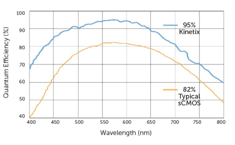
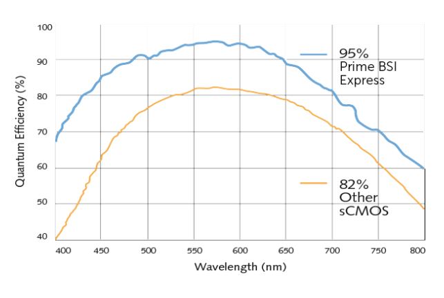
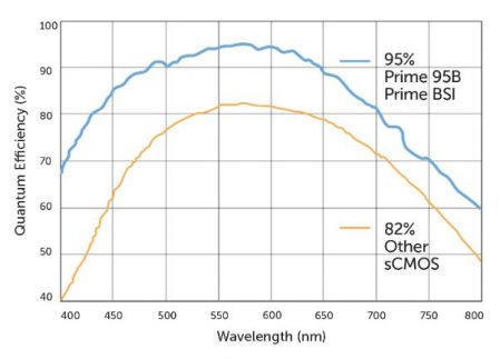
The Kinetix back-illuminated sCMOS camera achieves a near-perfect 95% quantum efficiency. By bringing the light in from the back of the sensor, photons land directly onto the light receiving surface, maximizing light collecting capability. The Kinetix combines 95% quantum efficiency with a low 1.2 e- read noise to deliver the most sensitive sCMOS camera at over 400 frames per second.
High Resolution
The Kinetix features 6.5 µm x 6.5 µm pixels, the accepted standard for most live cell applications using 40x and 60x magnification. This pixel size provides highly detailed images across the imaging plane and is most suitable for the broadest range of microscope objectives.
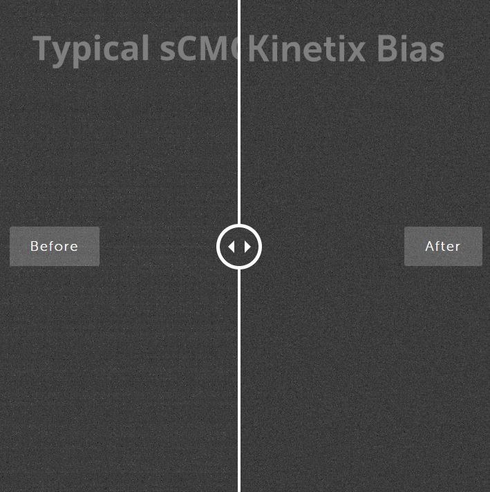
Superior Background Quality
The Kinetix features Pattern Noise Reduction Technology and Correlated Noise Reduction Technology to ensure that it delivers clean, pattern-free images with minimal pixel defects, delivering improved image quality in low light conditions.
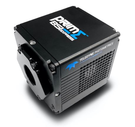
Prime BSI Express Scientific CMOS(sCMOS)는 최적화된 컴팩트 플랫폼으로 최적화된 Pixel 디자인, USB 3.2 Gen 2 연결성, 완벽한 95% Quantum Efficiency를 통해 고해상도 고감도 이미징의 완벽한 균형을 제공함으로써 최대한의 신호를 감지합니다.
UV(200-400nm), Visible(400-700nm), Near Infrared(700-1000nm)
Read Noise(median)
1.0 e-
Sensor Area
13.3mm X 13.3mm
Frame Rate
43fps @ 16bit / 43fps @12bit / 95fps @11bit
Prime BSI Express
4.2M pixels (2048×2048)
Sensitivity
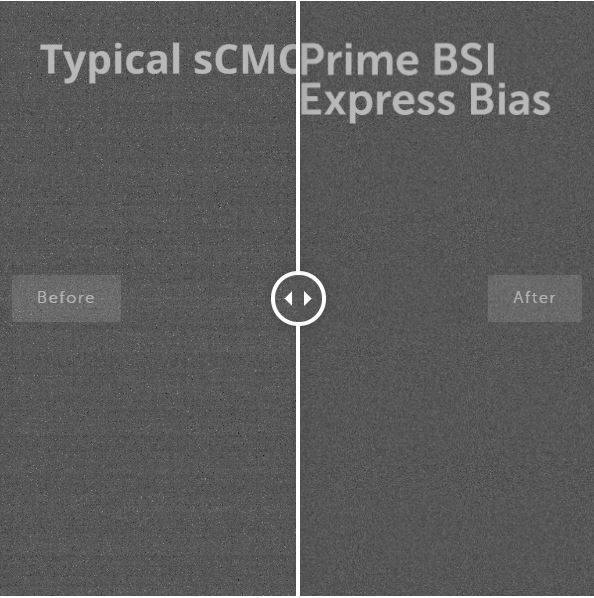
By bringing the light in from the back of the sensor, photons land directly onto the light receiving surface, maximizing light collecting capability. The Prime BSI Express combines 95% quantum efficiency with the low 1.0 e- read noise CMS mode to deliver the most sensitive camera based on sCMOS technology at over 40 frames per second.
Compact Form Factor
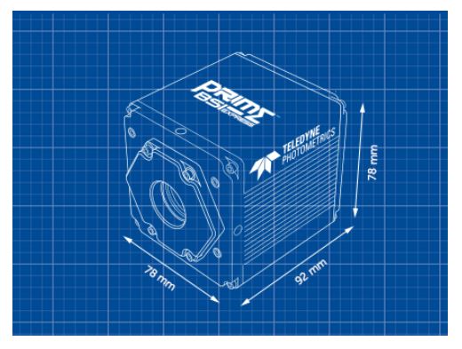
The Prime BSI Express is a compact 78mm x 78mm x 90mm with optimized cooling for the size, ideal for integration into new or existing configurations.
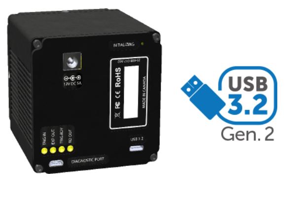
Interface and Integrate
Interfacing and integration is simple with the Prime BSI Express, which uses USB 3.2 Gen 2 for truly uncomplicated connectivity. Our standard PVCAM driver can be easily installed in Windows operating systems to get the Prime BSI Express running in minutes.
Resolution and Pixel Size
The Prime BSI Express features 6.5 µm pixels, the accepted standard for most live cell applications. This pixel size provides highly detailed images across the imaging plane and is most suitable for the broadest range of magnifications.
Superior Background Quality
The Prime BSI Express features Pattern Noise Reduction Technology and Correlated Noise Reduction Technology to ensure that it delivers clean, pattern-free images with minimal pixel defects, delivering improved image quality in low light conditions.
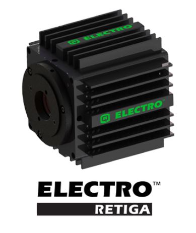
The advanced technical features of the Retiga ELECTRO were designed to empower electrophysiologists to image without exogenous noise that pollute recordings. This is accomplished by coupling regulated fanless cooling and external grounding with FPGA-based intelligent features to correct defective pixels.
High Sensitivity
The Retiga ELECTRO features 75% peak quantum efficiency (QE) and maintains a high QE across commonly used wavelengths in electrophysiology imaging. This is combined with low noise electronics to reveal weakest fluorescence signals. The Retiga ELECTRO also has an exceptionally low dark current so long stare applications will not suffer in image quality.
Simple Installation
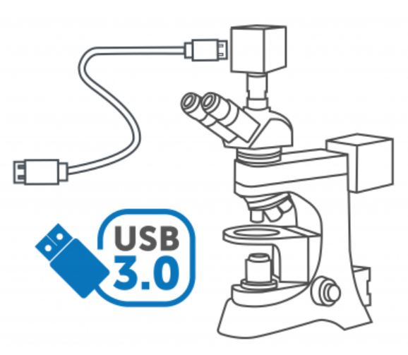
The Retiga ELECTRO delivers data over the simple, versatile and high-speed USB 3.0 interface. This standard, accepted interface is present on most modern computers, simplifying connectivity. The Retiga ELECTRO is also supported on the latest Windows operating systems ensuring modern system compatibility.
● USB 3.0:50MHz high frame rate를 적용하여 이미지의 끊김 없이 포커스 조절이 가능하며, 빠르게 원하는 샘플을 찾을 수 있음
● Real-time FPGA의 알고리즘을 적용하여 깨끗한 영상 구현
<Application>
■ Live cell Imaging and Fixed cell detecton
■ Time Lapse Imaging
■ Tile-and Stitch Microscopy
■ Stereo Microscopy
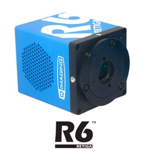
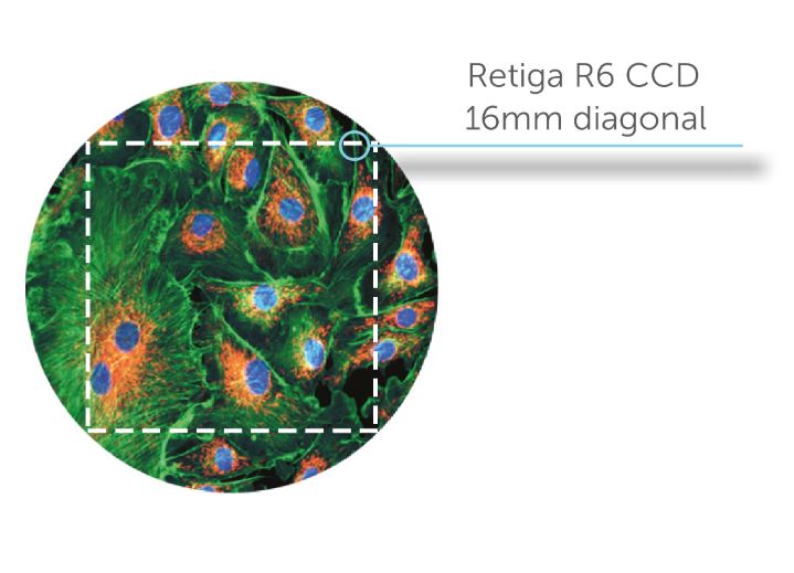
Large Field Of View
The Retiga R6 delivers a large, 16 mm diagonal field of view designed to increase throughput and maximize the amount of data captured in a single frame. Image larger samples or increase throughput by imaging multiple cells at once by taking advantage of a larger field of view.
High Resolution
The Retiga R6 features 6 million, 4.54 µm pixels to maximize the resolving power of your system. The small pixels provide highly detailed images across the imaging plane, which allows for the highest resolution when using lower magnification objectives.
Simple Installation
The Retiga R6 delivers data over the simple, versatile and high-speed USB 3.0 interface. This standard, accepted interface is present on most modern computers, simplifying connectivity. The Retiga R6 is also supported on the latest Windows operating systems ensuring modern system compatibility.
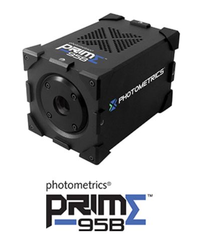
The Prime BSI back-illuminated sCMOS camera achieves a near-perfect 95% quantum efficiency. By bringing the light in from the back of the sensor, photons land directly onto the light receiving surface, maximizing light collecting capability. The Prime BSI combines 95% quantum efficiency with a low 1.0 e- read noise to deliver the most sensitive camera based on sCMOS technology at over 40 frames per second.
Resolution and Pixel Size
The Prime BSI features 6.5 μm pixels, the accepted standard for most live cell applications using 40x and 60x magnification. This pixel size provides highly detailed images across the imaging plane and is most suitable for the broadest range of microscope objectives.
Superior Background Quality
The Prime BSI features Pattern Noise Reduction Technology and Correlated Noise Reduction Technology to ensure that it delivers clean, pattern free images with minimal pixel defects, delivering improved image quality in low light conditions.
Advanced Triggering
Programmable Scan Mode provides increased control over the rolling shutter exposure and read-out functionality of CMOS sensors for applications that require control over the line time. Sequenced Multiple Acquisition Real Time Streaming (SMART) Streaming enables the Prime BSI to capture a continuous sequence of images while cycling through 16 pre-programmed exposure time values, delivering the fastest multi-channel acquisition times.
Advanced Features
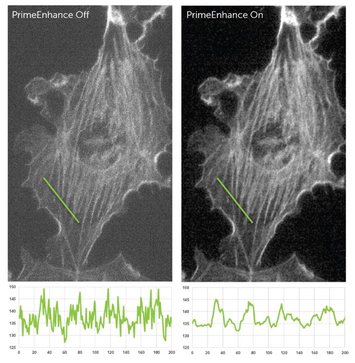
The Prime BSI offers PrimeEnhance active denoising which provides a real-time quantitative increase in the signal to noise ratio to maximize sensitivity at ultra-low light levels.