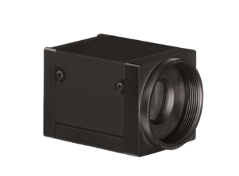
AcquCAM 23GR

AcquCAM 23GR


(Down load Catalog  – ACQUCAM 23GR)


  • USB 3.0 Color Camera
  • 1/1.2 ” Exmor Sensor
  • 1920×1200 pixel
  • Trigger input and I/O
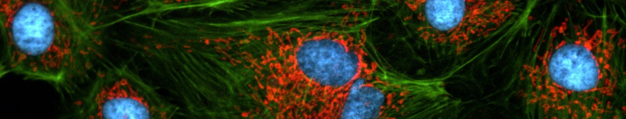
  • Available  of taking images with a wavelength of 650nm or more, such as CY5
  • High Sensitivity Sensor (Best)
  • Fast, seamless image transfer
  • Available of images with a very wide field of view
  • Low Noise Sensor


The displays live digital images with gradual smoothness and combines exceptional resolution  with faithful color reproduction.The new sensors feature a global shutter function and able to capture a high-speed moving image without focal plane distortion. High-speed processing, low noise and low power dissipation by using column-parallel A/D conversion. equipped with trigger mode, and the external pulse can control accumulation time. The Sensor also have a pulse output function to indicate respective conditions during shutter operation and can be coordinated with peripheral circuits. High-definition images can be displayed live at a rate of high frames per second. without compression. Such imaging quality enables even the finest cellular regions to be observed clearly and distinctly without deterioration. While focusing is made stress free.

Images with AcquCAM 23GR


Sample : bovine pulmonary artery endothelial cell, BPAEC
Filter Set : JNO-U(B), B(B), G(B)
Camera : AcquCAM 23GR2
C-mount adapter : 1X
Lens : UPLNAPO40X
Above three images are merged and processed using ARM S/W


JNO-ARM

Advance Realtime Monitor. Easy-to-Use, Image Enhancement, etc


1. Information

ARM is image analysis software for JNOPTIC AcquCAM cameras. This S/W is interchangeable with all WDM cameras regardless of camera brand and model and even more the most strength point is simple and easy use of length, area, angle, etc.


2. Simple measurement tools

  • Measurement Tools: Count, Distance, Angle, Area, etc.
  • Guide Lines: Cross Line, Rectangular Lattice, Circular Grid
  • Scale Panel on Live/Image Screen
  • Available measurement on LIVE image and saved image mode


3-1. Specialization for observation of fluorescence images

Improvement Function about Fluorescence Microscopy Image. (Used function: Auto Level, Low level, Pseudo Color, Merge Image)

Bottom on the left is merged image without improvement of image. And the bottom on the right is merged image after improvement of image. These images are shot by JNOPTIC AcquCAM Pro/G3 camera


3-2-1 Effect of high-contrast image with simple operation (Monochrome)

Improvement Function about Microscopy Image Monochrome. (Used function: Auto Level)

Effective improvement of image with simple operation. This image is shot by JNOPTIC Pro/S3 camera


3-2-2 Effect of high-contrast image with simple operation (Color)

Improvement Function about Microscopy Image. (Used function: Auto Level)

Effective improvement of image with simple operation. These images are shot by JNOPTIC AcquCAM Pro/G3 camera


3-3-1 Live Pseudo Color (for color camera)

When you get fluorescence images using color camera, if the unwanted channel was shown because of cross talk, you could remove this channel to take advantage of function of Live Pseudo.
Improvement Function about Fluorescence Microscopy Live Camera Image. (Used function: Live Pseudo Color)
 The extraction of wanted color information from color image (Remove BR on RGB source) These images are shot by JNOPTIC AcquCAM Pro/G3 camera

3-3-2 Live Pseudo Color (for Monochrome camera)

When you get fluorescence images using monochrome camera, you can raise efficiency of acquisition for fluorescence images to add similar false color to the color to be shown on the eyepiece in real time.
Improvement Function about Fluorescence Microscopy Live Monochrome Camera Image. (Used function: Live Pseudo Color)
 Improvement of usage environment for monochrome camera to add false color on the image. These images are shot by JNOPTIC AcquCAM Pro/S3 camera

4. JNO-MHU(Option Unit)

Available measuring height with additional unit, JNO-MHU

※ JNO-MHU is sold separately as optional unit.

Left: Focus on top of sample (Z-axis reset), Right: Focus on bottom of sample (Z-axis height measurement)

 < Measuring condition : over 20x objective, temp 20°C >

Responding Model Measurement value unit Measurable height Error of measuring 1000㎛ Recommended measuring height
CX 41 0.2 ㎛ -9.9 ~ 29.9㎜ Below ±20㎛ Below ± 2000㎛
CKX 41 0.2 ㎛ -9.9 ~ 29.9㎜ Below ±20㎛ Below ± 2000㎛
BX – FM 0.2 ㎛ -9.9 ~ 29.9㎜ Below ±20㎛ Below ± 2000㎛
BX 51/53 0.1 ㎛ -9.9 ~ 29.9㎜ Below ±10㎛ Below ± 1000㎛
MX 51 0.1 ㎛ -9.9 ~ 29.9㎜ Below ±10㎛ Below ± 1000㎛
MX 61L/61 0.1 ㎛ -9.9 ~ 29.9㎜ Below ±10㎛ Below ± 1000㎛


5. Reporting to Excel


Specification

Spec Description
Size 1/1.2 inch
Resolution H: 1920, V: 1200
Pixel size H: 5.86 µm, V: 5.86 µm
Video formats& Frame rate 1920×1200 RGB24@ 54 fps
1920×1200 Y800@ 162 fps
1920×1200 Y16@ 80 fps                             1920×1200 Y411 @ 108 fps
1920×1200 YUY2 @ 81 fps
Dynamic range 8 / 12 bit
Exposure time 20 ㎲ to 30s
Lens mount C/Cs mount
Interface USB 3.0
Power supply 4.5 to 5.5 VDC
Dimensions H: 29 mm, W: 29 mm, L: 43 mm
Mass 65g


More images with AcquCAM 23GR

-> For more details, click here.