카테고리 보관물: Bio Microscope

BX41-Bio

BX41

연구용 정립 현미경

BX41은 의학분야를 위한 맞춤형 장비입니다. 강화된 본체, 진보된 광학계를 가진 BX41은 다양한 용도와 인체공학적인 편리함을 제공합니다.

인체공학적 디자인(효율성 및 편리성)

  • Y-모양의 본체는 사용자가 다루기 쉬운 형태로 되어 있으며 운용의 안정성을 제공합니다.
  • 낮은 위치에서 초점을 조절 할 수 있는 본체와 앞쪽에서 조절이 가능한 조명장치, 조동나사 압력 조절, 퀠러조명, 탈․부착 가능한 nosepiece로 구성되어 있어서 사용자의 검경자세를 최대한 편안하게 만들어드립니다.
  • 높은 정확도와 깔끔한 디자인의 제물대는 X, Y 움직임을 사용자에 맞게 조절 가능하며 왼손/오른손잡이 선택도 가능 합니다.

유연성 있는 형태의 시스템

  • 광학계의 변동없이 다른 액세서리를 부착 가능 합니다; 다양한 video 장비, 필름 카메라, 디지털 카메라가 장착 가능합니다. 새롭게 개발된 BX-URA2와 BX-RFA 장착시 보다 효율적인 형광 input/output을 제공합니다.
  • BX41은 UIS2 광학계가 적용된 모델로서 밝고, 깨끗하고, 고대비의 결과물을 만들어줍니다.
  • 6V/30W 할로겐 광원의 프리셋 버튼 이용하면 최적의 색온도 가지는 밝기를 만들 수 있습니다.
  • 8-position 콘덴서에는 Brightfield, Darkfield , Phase Contrast , polarization, and DIC (Nomarski)등과 같은 다양한 필터를 장착할 수 있습니다. 또한 1.25X-100X를 콘덴서 변환 없이 관찰이 가능 합니다.


IX81

IX81

연구용 전동 현미경

IX81은 올림푸스의 최상급 도립현미경 시스템입니다.

IX81의 모듈형 본체와 광학계는 최대 9개까지의 input/output 장비를 장착 할 수 있도록 디자인 되었습니다. 파트 조합에 따라 4개의 port에서 동시에 이미지를 얻을 수도 있습니다. 내장된 Z-축 조절 모터, 6-position nosepiece와 광로변환장치를 내장한 IX81은 다양한 파장의 형광이미지를 밝고 선명하게 얻을 수 있습니다.

Highlights

IX81은 독특한 ‘Two-tiered and V-shaped optical design’을 채용하여 광학계 상의 빛 손실을 최소화 하였으며, 본체의 수리/변형 없이 다양한 device port를 이용하여 깨끗하고 밝은 이미지를 얻을 수 있습니다.

올림푸스의 ‘Two-tiered optical system’은 광학계와 기계적 변형 없이 여러 방향으로의 접근이 가능 합니다. 뒤쪽 port는 CCD 카메라 뿐만 아니라 다른 특수 장비(레이저 스캐닝 유닛 등)도 쉽게 장착할 수 있습니다. 현미경 본체는 자동화 된 6-position nosepiece, 광로변환스위치, 0.01um로 조절 가능한 Z축 모터가 장착 되어있습니다. 또한 본체의 디자인과 강도 높은 알루미늄 소재는 안정성과, 견고함을 보장해 드립니다. 또한 새롭게 디자인된 nosepiece로 하여금 더욱 높은 안정성을 가지게 되었습니다.

  • 부가적인 전동 액세서리 장치로는 6-position motorized condenser, 6-position fluorescence filter turret, transmitted and reflected light shutters, filter wheels 등이 있습니다.
  • IX81의 6-position fluorescence filter turret은 DIC/polarize 및 형광관찰 시의 필터변환이 컴퓨터로 조절되며 filter wheel과 전동 shutter를 동시에 작동할 수 있습니다. 이는 기존의 수동으로 하던 복잡한 조작을 클릭 한 번으로 수행할 수 있음을 의미합니다.
  • 자동화 기능, 할로겐 및 형광 광원은 발열 및 전기적 잡음을 발생시킵니다. 이를 제거하기 위해 파워 장치와 조절 장치는 현미경 본체와 분리되어 있습니다.
  • 새롭게 개발 된 IX2 현미경은 dual camera adapter를 left side port와 low, upper, rear port에 장착하여 동시에 여러 이미지를 얻을 수 있습니다. 또한 이러한 포트에 장착되는 dual adapter에는 dichromatic mirror를 장착되어있으므로 서로 다른 파장의 이미지를 동시에 얻음과 동시에 실시간 ratio-metric imaging이 가능합니다.
  • 모든 현미경 조작부는 인체공학적으로 제작 되었으며, 진동 없는 조절이 가능합니다. 또한 본체와 핸드스위치에 사용자가 원하는 기능을 별도 입력 할 수도 있습니다.


Frame Construction

  • 뛰어난 본체의 안정과 항진동 설계는 patch clamping과 같은 관찰에 최적화 되어 있습니다. 내장된 Z-축 모터는 0.01µm 씩 조절이 가능하여 기본기능 뿐만 아니라 IX81을 기반으로한 시스템 장비 구축에 이상적인 현미경 프레임입니다.
  • 4개의 Imaging port를 포함한 9개의 port는 뛰어난 확장성을 제공합니다. 다수의 CCD 카메라를 장착하면 real time co-localization imaging 및 spectral un-mixin이 가능 합니다.
  • 본체는 컴팩트하게 디자인 되어있으며 MMC(Multi-Metal Complex)를 소재로 사용하여 견고성과 항온성을 제공합니다.
  • 새로 구성된 Nosepiece focus mechanism은 조작거리를 단축하여 장시간의 샘플관찰에 안정성을 제공합니다.
  • 외부장치를 장착하기 쉽게 디자인 되었습니다.
  • 방수처리된 nosepiece guide는 이물질이 광학계 내부로 유입되는 것을 방지해 줍니다; 방진/방습 디자인은 내부 광학계 표면부식을 막아 줍니다.


Deconvolution / Laser scanning confocal

  • 10nm의 고정밀 Z-축 모터는 Confocal Imaging 또는 Deconvolution Imaging과 같은 optical sectioning에 강력한 기반을 제공합니다.
  • 본체는 무진동 테이블과 쉽게 결합 및 고정이 가능합니다.
  • FluoView 1000 Confocal scan unit과 IX81을 결합시킨 상태에서 현미경의 양쪽에 다른 장비들을 장착 시킬 수 있습니다. 물론 연구자가 원하는 다른 port로도 Confocal scan unit을 장착 하실 수 있습니다.
  • 최소화/안정화에 중점을 둔 디자인과 외부 전원, 그리고 더욱 견고해진 focus mechanism은 Digital imaging을 위해 최적화 되어 있습니다.

Optical Features and Optical Efficiency

  • Single reflection방식인 V-shaped optical path는 보다 밝고 깨끗한 이미지를 제공합니다.
  • Binocular와 tilting binocular, 그리고 trinocular tube가 장착 가능합니다.
  • 본체는 1.6X (optional 2x) 배율 변환기를 기본 장착되어, 육안관찰 시 기본 배율을 뛰어넘는 관찰이 가능합니다. (카메라 이미지 시에는 적용이 되지 않습니다.)
  • 본체의 bottom port는 극단적으로 적은 양의 빛을 검출하는 실험 사용하실 수 있습니다. (특수 제작된 재진대가 필요합니다.)

Fluorescence

  • 개선된 광학계는 고배율렌즈에서 약 20% 정도의 밝기 증가를 이루었습니다. 이는 낮은 조도에서도 높은 신호대잡음비를 만들 수 있음을 말해줍니다.
  • 새롭게 고안된 L-Shaped 광로는 형광 광원의 이상적인 centering을 가능하게합니다.


BX61-Bio

BX61

연구용 전동 현미경

BX61은 소프트웨어 제어가 가능한 전자동 시스템입니다.
포커스조절, 광원제어, 렌즈 및 필터교환 등의 핵심기능을
Software setting에 저장하여 연구자가 요구하는 조작들을
쉽게 불러내어 사용할 수 있습니다.
BX61은 다음과 같은 작업에 최적화 되어 있습니다.

  • 반복적인 작업
  • 복잡한 조작을 요구하는 Time-lapse 실험
  • 인터넷 또는 네트워크를 통한 원격제어
  • 시료의 오염을 최소화해야하는 작업

BX61의 핵심은 내부에 장착된 포커싱모터입니다.
이를 통해 최소 0.01 micron 단위로 조절이 가능하므로
Z-축으로의 이동 및 측정을 보다 정확하게 할 수 있으며,
주변 공간을 효율적으로 사용할 수 있도록 소형화/간편화되어있습니다.
또한, 현미경에 발생할 수 있는 열전도를 최소화하였으며,
전기적간섭도 방지되어 실험을 정확하게 수행 할 수 있습니다.

또한 BX61은 여러 목적의 실험에 맞는 액세서리들을 장착할 수 있습니다. 이러한 액세서리들은 통합 컨트롤러인 BX-UCB와 컴퓨터를 통하여 간편하게 제어가 가능합니다.
여러가지 액세서리 조합으로 다음과 같은 기능 수행이 가능합니다.

  • 여러 종류의 렌즈를 장착 할 수 있는 6-position nosepieces 조절
  • 6~8개의 형광 필터 터렛과 형광광원을 open/close하는 shutter 조절
  • 8가지의 콘덴서 조합 가능
  • DIC, Phase contrst(위상차), Darkfield, Polarize 관찰
8-position 콘덴서
(DIC, 위상차, Darkfield, 편광 관찰 가능)
7-position 대물레즈
nosepiece를 이용 다양한 렌즈 장착 가능

이 외에도 Auto focus, stage control등과 같은 다양한 기능들을 제어 하실 수 있습니다.


CKX31/CKX41

CKX31/CKX41

실험용 도립 현미경

CKX현미경은 UIS2 광학계로 구성되어 밝고 선명하고 편리한 조작성을 가지고 있기에 세포 관찰에 적합합니다.

CKX31은 발생학, 면역학 그리고 생리학 등 다양한 생물학 분야에서 사용 가능하며 뛰어난 가격대비성능을 가지고 있습니다.

GFP관찰을 위한 형광광원장치를 구매, 장착하면 한 단계 발전된 형광 영상을 얻을 수 있습니다. 디지털 이미징과 모니터링을 위한 디지털 카메라 장비도 장착 가능합니다. CKX41은 petri-dish, culture flasks, multi-well plate 등을 이용하여 세포의 viability 관찰이 가능합니다.

  • CKX31 & CKX41은 UIS2 광학계로 디자인 되어 있습니다.
  • 본체가 작으므로 클린벤치와 같은 협소한 공간에서도 활용이 가능합니다.
  • 위상차 및 새 Relief Contrast 관찰을 위해 기본적인 광학능력이 개선되었습니다.
  • A pre-centered slider를 사용하였으므로 별도의 얼라이닝 작업 없이 위상차 관찰이 가능합니다.
  • 대안렌즈의 상하 각도 조절이 가능한 경통은 다양한 환경에서도 편안한 관찰각도를 만들어 줍니다. (CKX41에 부가 장착 가능)
  • 강력한 6V/30W 할로겐램프는 밝고 선명한 세포관찰을 위해 풍부한 광원을 제공합니다.
  • 유리로 제작된 제물대 삽입 판(Glass plate)은 대물렌즈의배율을 쉽게 구별할 수 있도록 해줍니다.(CKX41에 부가 장착 가능)
  • 형광광원의 특징; Blue, Green excitation cube를 위한 dual slider 그리고 상대적으로 가격이 저렴한 50W 수은램프를 제공 합니다. (CKX41에 부가 장착 가능)
  • 위상차 조절이 필요 없습니다. (CKX31 일반모델 적용)


IX71

IX71

연구용 도립 현미경

IX2 series의 IX71 도립 연구 현미경은 보다 복잡하고 어려운 실험 분야에 대응할수 있도록 설계되었습니다.

시스템 장비인 IX71은 9개의 확장 포트에 다양한 장비를 장착할 수 있습니다. 4개(이상)의 포트에 영상장치를 장착하여 4종류의 이미지 동시 촬영이 가능합니다. 또한 개선된 형광기술이 적용되어 다양한 파장의 형광샘플을 관찰할 수 있습니다.

Highlights

IX71은 Two-tiered, V-shape광학 디자인으로 엄청난 확장성을 제공하며 빛의 손실을 최소화하였기 때문에 더욱 밝고 깨끗한 이미지 관찰이 가능합니다.

OLYMPUS의 two-tiered 광학계는 제물대 및 형광 광로의 변형 없이 Upper, Lower port를 이용하여 샘플 영상을 얻을 수 있습니다. Rear port를 이용하면 보다 넓은 작업공간을 확보할 수 있습니다.

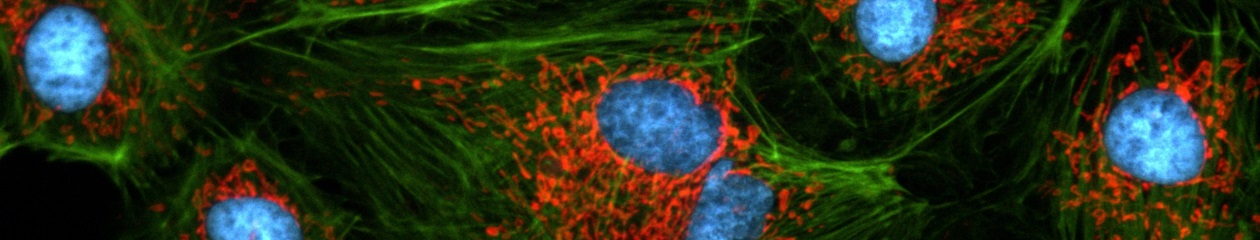
IX71에 dual camera adapter를 추가한 뒤두 대의 CCD카메라를 장착하면 여러 이미지를 동시에 얻을 수 있습니다. 또한 이 어댑터에 형광파장분리장치를 장착하면 두 가지의 형광파장을 동시에 관찰할 수 있습니다.

열에 대한 안정성을 확보하기 위해서 본체와 전원장치가 분리되어있습니다. 또한 현미경 프레임은 Cast ADC-12 aluminum 재질이 사용되었고 정밀한 컴퓨터 제작을 통해 제작되었습니다. 새롭게 디자인된 nosepiece는 장시간 관찰에서도 신뢰성있는 결과물을 만들어줍니다.

빛 손실이 최소화 된 V-light path와 개선된 apochromatic relay lens는 밝고 선명한 이미지를 만들어냅니다.

견고함을 바탕으로 한 인체공학적 설계는 안정성과 편의성을 제공합니다.

3.7mm Working distance, 0.9 NA, patch clamp 부착이 용이한 40°의 각도를 가진 Water immersion lens 콘덴서도 선택할 수 있습니다.

Straight 또는 L-Shaped 형광 광원장치는 340nm의 높은 UV 투과 능력을 가지고 있으며 6-position filter cube turret과 조합하여 밝은 형광 이미지를 얻을 수 있습니다.


Frame Construction

뛰어난 본체 안정성과 진동 없는 조작성은 Patch clamping과 같은 어플리케이션에 적합합니다.

컴퓨터 디자인된 본체는 ADC-12 aluminum로 제작되어 온도변화에 안정하며 외부 전원장치를 사용함으로 안정성은 배가 됩니다.
새로운 nosepiece 작동 부위의 조작 거리가 더 짧아 졌기에 장시간의 관찰에도 초점흐름이 최소화됩니다.
본체 디자인은 여러 가지의 장비를 장착하기 편하도록 Y-shape로 제작이 되었습니다.


Equipment

뛰어난 본체 안정성과 진동 없는 조작성은 Patch clamping과 어플리케이션에 적합합니다.

컴퓨터 디자인된 본체는 ADC-12 aluminum로 제작되었으며 온도변화 안정성 확보를 위해 전원을 외부에 장착 하였습니다.
새로운 nosepiece는 작동 거리가 더 짧아 졌기에 장시간 샘플을 관찰할 때 초점흐름 현상이 최소화됩니다.
본체 디자인은 여러 가지의 장비를 장착하기 편하도록 Y-shape로 제작이 되었습니다.

  • 방수 nosepiece 보호 장비를 사용하면 시약이나 배지와 같은 액체를 쏟을 경우 현미경 내부의 광학계를 보호할 수 있습니다. ; 방진/방습 디자인은 광학계의 표면의 더러워짐을 방지해 줍니다.
  • Patchclamping/Electrophysiology(전기생리학)pre-tapped mount가 내장된 본체는 다른 액세서리나 micromanipulator를 쉽게 장착할 수 있습니다.
  • 높은 NA(0.9 NA)를 가지는 Water Immersion/DIC 콘덴서 시스템은 patch clamping을 위한 3.7mm의 working distance와 40°의 각도로 pipette 사용을 용이하게 해줍니다.
  • 새롭게 디자인된 콘덴서는 진동을 줄이기 위해 Click-stop 방식으로 설계되었습니다.
  • 현미경의 본체와 제물대는 Grounding electrode를 장착하실 수 있도록 특성화 되어 있습니다.

Illumination Columns

  • 2개의 12V/100W 광원장치를 rear port로 연결하고 뒷면으로 젖히시면 manipulator의 샘플 접근 영역을 확보할 수 있습니다. 또한 콘덴서도 독립적으로 조절 가능하여 위쪽으로 젖혀 놓을 수 있습니다.
  • 100W 광원 장치에 신속한 조절이 가능한 교환식콘덴서를 장착할 수 있습니다.
  • 100W 콘덴서는 relief contrast를 위해 이동 거리가 넓어 졌습니다.
  • 또한 저렴한 비용의 6V/30W광원 장치를 사용하실 수 있습니다.

Optical Features and Optical Efficiency

  • Two-tiered 광학 디자인으로 제작된 본체는 최대 9개의 광학장비 장착이 가능합니다.
  • Up to 4 ports can have simultaneous access to a primary image allowing high quality, high speed, chromatically separated imaging
  • Two rear ports are available without modifying the position of the stage, fluorescence illuminator or nosepiece assembly
  • 단일 반사, V-shape 광학 경로는 apochromat relay lens를 통해 밝고 선명한 영상을 얻을 수 있습니다.
  • Binocular, tilting binocular 그리고 trinocular tube 사용 가능
  • 본체에 1.6X(option 2X)배율 교환기가 기본 장착되어 있으므로 별도 장치 없이 배율을 높일 수 있습니다.

Fluorescence

  • 개선된 straight 광원장치는 이전의 모델에 비해 광학요소의 개선을 통해 20% 더 밝은 이미지를 얻을 수 있습니다.
  • 새로운 L-Shaped 광원장치의 특성은 Aperture stop, Field Stop이 내장되어 쉽게 형광을 중앙으로 조절 하실 수 있습니다.


IX51

IX51

연구용 도립 현미경

IX51 도립현미경은 전문 연구소 및 임상 분야에 사용되는 현미경입니다. 모듈화 디자인, 뛰어난 광학 성능은 형광을 이용한 세포배양관찰에 적합합니다. 

Highlights

우수한 안정성과 인체공학설계는 다수의 사용자가 있는 연구실에서 별도의 얼라이닝 없이 장비사용이 가능합니다.

개선된 위상차 기술은 다양한 Well plate나 culture dish에서도 우수한 이미지를 얻을 수 있습니다.

부가적인 pre-centered phase optics는 위상차 관찰을 위한 얼라이닝이 필요없기에 검경에만 몰두하실 수 있도록 해줍니다.

현미경 내부는 ‘straight path’방식으로 설계되었기 때문에 밝은 형광이미지를 만들어 줍니다. 또한 여러 관찰법과 관련된 광원장치들을 6-position 필터휠안에 조합할 수 있습니다. L-형태의 광원장치를 이용하면 Aperture / Field stop을 더욱 쉽게 조절할 수 있습니다.

모듈화 디자인은 사용자가 필요한 시스템의 구성을 각각의 실험실에 맞게 구성할 수 있습니다. 또한 현미경 전용 비디오 장치나 디지털 카메라를 장착하게 되면 육안으로 영상을 관찰하면서 컴퓨터로 디지털 이미지를 얻을 수 있습니다.

  • 견고하고 편리 IX51 현미경은 빠르고 쉽고 정확한 조작이 가능하기에 정확한 검경이 가능합니다.
  • 제물대의 위치가 낮기 때문에 샘플을 올리고 교환하는 작업을 더욱 쉽게 할 수 있습니다.
  • 기본 제물대를 이용하셔도 모든 종류의 culture dish, flask, well plate를 사용하여 시료관찰이 가능합니다.
  • ‘Flex handle’ 제물대는 쉽고 빠른 조절이 가능합니다.
  • 대물렌즈 마운트에 방수 장비를 장착하면 샘플에 사용된 시약으로부터 내부 광학계를 보호할 수 있습니다. ; 방진/방습 디자인은 광학계 표면을 보호합니다.
  • Interchangeable observation tubes: centering telescope가 내장되어있으므로 별도의 장비없이 위상차 얼라이닝이 가능합니다. 삼안 경통은 카메라 장착이 가능합니다.
  • Side port를 위한 프리즘은 100%반사 또는 80%반사&20%투과 타입이있습니다. 실험목적에 따라 선택하실 수 있습니다.
  • 100W 할로겐 광원장치를 illumination pillar에 부착할 수 있습니다.

Complete and Modular Condenser System

4종의 집광기는 샘플과 배양용기의 종류에 따라 선택이 가능합니다.

  • 182mm의 working distance를 가진 Super Long Working Distance Condenser(SLWD)는 대형 배양 용기에 담긴 시료를 4X, 10X, 20X 대물렌즈로 관찰이 가능하게 합니다.
  • 73mm의 Long Working Distance Condenser를 이용하면 4X 부터 40X 대물렌즈에서 위상차영상을 관찰할 수 있습니다. 특히, 10X 부터 40X 까지 위상차 장치의 조절 없이 관찰할 수 있는 독특한 디자인으로 제작 되었으며 더 넓은 working distance를 확보하기 위해 Condenser 앞쪽 렌즈를 제거할 수도 있습니다.
  • Relief Contrast Condenser는 45mm의 Working distance와 0.5 NA를 가지며 플라스틱 용기에 담겨진 샘플로부터 3D relief image를 만들어 낼 수 있습니다. 특히 micro-injection 작업 시 Oocyte를 선명하게 관찰할 수 있습니다.


CX21

CX21

실험용 정립 현미경


CX21은 생물학 분야의 교육용 현미경으로서 사용과 유지보수가 편리한 것이 장점입니다.

  • 새로운 광학계가 채용 된 UIS2 Plan Achromat 렌즈(4x, 10x, 40x, 100x oil)는 주변부 화질 저하 없이 깨끗하고 선명한 이미지를 관찰할 수 있습니다.
  • 두 종류의 제품이 준비되어있으며 경제적인 가격이라 교육용도로 사용하기에 무리가 없습니다. (Package 1: 4x, 10x, 40x objectives; Package 2: 4x, 10x, 40x, 100x objectives)
  • F.N. 18 대안렌즈
  • 항 곰팡이 처리는 높은 습도에서도 광학계를 보호하기 때문에 단체 실습실과 같은 환경에서도 보관이 용이합니다.
  • 조작이 부드럽고 4개의 렌즈가 장착된 revolving nosepiece
  • 고정형 Abbe 콘덴서는 ‘Köhler illumination’방식으로 높은 N.A. 1.25을 제공해 줍니다.
  • 6V/20W 할로겐램프는 깨끗하고 안정적인 광원을 제공합니다. 내장된 aspherical collector lens는 모든 관찰 영역에 일정하고 안정한 빛을 제공합니다.
  • 이동이 용이한 디자인으로 교실 선반 같은 장소에도 보관이 가능 합니다.
  • 제물대는 조절 나사가 낮은 부위에 있으므로 편하게 X, Y 조작이 가능합니다.
  • 실습 용도에 맞추어 규격화되어 있으므로 유지/보수가 용이합니다.


CX33

CX33 Biological Microscope

Comfortable, High-Throughput Routine Microscopy

The CX33 microscopes enable users to remain comfortable during long periods of routine microscopy observations. The microscope frame conforms to the user’s hands and the location of the control knobs maximize ergonomics to improve work efficiency. Users can quickly set a specimen with one hand, while adjusting the focus and operating the stage with the other hand with minimal movement. The microscope also features an optional camera port for digital imaging.

Maintain Preferred Observation Conditions with Minimal Adjustments

Uniform Illumination with Consistent Color Temperature

The color temperature of the CX LED illumination produces daylight conditions, so specimens can be viewed with their natural colors. The color temperature is consistent at any brightness, so users don’t have to spend time making adjustments when they change brightness. The LEDs have a long 60,000-hour lifetime built into the design, helping reduce cost, and the brightness level remains stable throughout the LED’s life.

Excellent Optical Performance for Flat Images

The microscope employs Plan Achromat objectives, which provide clear images with high image flatness over a wide field of view. This helps users view specimens clearly and evenly illuminated during routine microscope observations.

Change Magnification without Adjusting the Condenser

 Users can change the magnification from 4X to 100X without moving the top lens on the condenser.

Remain Comfortable during Extended Usage

Single-Handed Sample Placement

A specimen can be quickly slid in and out with one hand. The specimen holder opens a little and firmly retains the specimen during operation. The versatile holder accommodates a variety of slide types, including a hemocytometer.

Smooth Magnification Change

The low-positioned revolving nosepiece enables users to quickly change magnifications with minimal arm movement between focusing, greatly improving work efficiency during prolonged use.

Ergonomically-Positioned Focus Knob

The low-positioned focusing knob enables users to make observations while keeping their hands and forearms rested on the desk, helping provide comfort. The focusing stopper prevents a specimen from accidentally hitting an objective when working under a high magnification.

Ergonomic Stage and Eyepiece Position

The low-positioned stage is designed to enhance comfort and reduce fatigue. The stage surface can be widely seen from the eye point position, which enables users to smoothly set and check specimens on the stage. The stage knob can be controlled with just a light touch and can be adjusted at the same time as the focusing knob, since they are located close together.

Specimen Holders that Match Your Observation Style

Stage accessories improve efficiency when users need to observe a large number of specimens. With the specimen holder sheet, a specimen can be freely operated by a finger on the sheet and can be precisely adjusted using the stage knob. The double specimen holder can retain a large specimen or two specimens.


CX43

CX43 Biological Microscope

Comfortable, High-Throughput Routine Microscopy

The CX43 microscope enable users to remain comfortable during long periods of routine microscopy observations. The microscope frame conforms to the user’s hands and the location of the control knobs maximize ergonomics to improve work efficiency. Users can quickly set a specimen with one hand, while adjusting the focus and operating the stage with the other hand with minimal movement. The microscope also features an optional camera port for digital imaging.

Maintain Preferred Observation Conditions with Minimal Adjustments

Uniform Illumination with Consistent Color Temperature

The color temperature of the CX LED illumination produces daylight conditions, so specimens can be viewed with their natural colors. The color temperature is consistent at any brightness, so users don’t have to spend time making adjustments when they change brightness. The LEDs have a long 60,000-hour lifetime built into the design, helping reduce cost, and the brightness level remains stable throughout the LED’s life.

Excellent Optical Performance for Flat Images

The microscope employs Plan Achromat objectives, which provide clear images with high image flatness over a wide field of view. This helps users view specimens clearly and evenly illuminated during routine microscope observations.

Select and Set Your Contrast Level

Users can preserve their favorite contrast by locking the aperture diaphragm. It stays fixed at the optimally chosen position if it is accidentally touched while changing slides.

Change Magnification without Adjusting the Condenser

 Users can change the magnification from 4X to 100X without moving the top lens on the condenser. 2X magnification is also available by simply setting the objective and the condenser turret to 2X position.

Simple Fluorescence Observation

Fluorescence observation is simple and easy. Plug the compact fluorescent illuminator into the microscope frame for fluorescence observation. Its LED light source is pre-centered, and the transmitted illumination is shuttered by simply setting the condenser turret to the FL position. This reduces background noise in the fluorescence image from incidental light coming from the top lens of the condenser.


Remain Comfortable during Extended Usage

Single-Handed Sample Placement

A specimen can be quickly slid in and out with one hand. The specimen holder opens a little and firmly retains the specimen during operation. The versatile holder accommodates a variety of slide types, including a hemocytometer.

Smooth Magnification Change

The low-positioned revolving nosepiece enables users to quickly change magnifications with minimal arm movement between focusing, greatly improving work efficiency during prolonged use.

Use Up to Five Objectives

For added flexibility, up to five objectives can be supported by the revolving nosepiece. In addition to general objectives, users can select a 2X objective for wide area observation or objectives for phase contrast. These objectives with long working distances help keep specimens from getting damaged.

Ergonomically-Positioned Focus Knob

The low-positioned focusing knob enables users to make observations while keeping their hands and forearms rested on the desk, helping provide comfort. The focusing stopper prevents a specimen from accidentally hitting an objective when working under a high magnification.

Ergonomic Stage and Eyepiece Position

 The low-positioned stage is designed to enhance comfort and reduce fatigue. The stage surface can be widely seen from the eye point position, which enables users to smoothly set and check specimens on the stage. The stage knob can be controlled with just a light touch and can be adjusted at the same time as the focusing knob, since they are located close together.

Specimen Holders that Match Your Observation Style

Stage accessories improve efficiency when users need to observe a large number of specimens. With the specimen holder sheet, a specimen can be freely operated by a finger on the sheet and can be precisely adjusted using the stage knob. The double specimen holder can retain a large specimen or two specimens.

Simplified Fluorescence Observation

Fluorescence observation can be easily set up on the standard configuration while keeping the eye point the same as other observation methods. Simply plug the compact fluorescent illuminator into the back of the microscope frame.

Versatile Applications

The universal condenser offers a variety of observation methods and future upgradability. In combination with the five-position revolving nosepiece, multiple applications can be covered using the single microscope frame.

Brightfield

Brightfield

Phase Contrast

Fluorescence


Accessories

Simple polarizing intermediate attachment/CX3-KPA

Offers polarized observation of urate crystals and amyloid in combination with apolarizer and analyzer.

Eyepoint adjuster/ U-EPA2

Raise the eyepoint position by 30 mm for added comfort.

Arrow pointer/ U-APT

Insert an LED arrow into your image; great for digital imaging and presentations.

Dual observation attachment/U-DO3

Enables dual, simultaneous observation of a single specimen from the same direction with equal magnification and brightness for both operators. A pointer can be used to indicate specific sections of the specimen to simplify the training process and enhance discussion.


BX53-P

BX53-P 정립 현미경

편광 현미경

Olympus BX53-P 편광 현미경은 UIS2의 무한보정 광학계와 독자적인 광학 설계의 조합을 편광 어플리케이션에서 뛰어난 성능을 제공합니다. 폭 넓은 컴펜세이터의 라인업은 BX53-P를 어떤 분야에서의 관찰과 측정실험도 가능하도록 만들어줍니다.

UIS2 광학계는 뛰어난 확장성을 제공합니다.

무한 보정의 장점을 최대화하여, UIS2 광학계는 광학 현미경 성능의 저하를 막고, 분석판(Analyzer), 틴트 플레이트(Tint Plate)나 컴펜세이터(Compensator)가 광로 상에 있어도 배율 변화가 없습니다. 높은 수준의 시스템 유연성 유지를 위해, BX53-P는 BX3 시리즈 시스템 현미경의 중간 연결 장치(Intermediate attachment), 카메라 및 이미징 시스템과도 호환됩니다.


편광 특성의 개선

ACHN-P와 UPLFLN-P에 사용된 정교한 디자인과 제조 기술은 내부 변형을 줄여 줍니다. BX53-P 편광 현미경은 EF 값이 높은 Filter가 적용된 편광판(Polarizer)과 분석판(Analyzer)를 사용하여 영상의 대비가 뛰어납니다. 연구의 다양성과 어플리케이션의 요구들을 충족하기 위한 범용 UPLFLN-P 시리즈 대물렌즈는 미분간섭(Nomarski DIC) 및 형광현미경, 편광 관찰에 이르기까지 많은 관찰법들을 폭넓게 수용하도록 설계되어왔습니다.
* 흡광인자(EF_Extinction Factor)는 평행과 교차편광 필터 사이의 휘도비(Brightness Ratio)를 의미합니다. 흡광인자의 값이 클수록 흡광도(Extinction)가 높습니다.


컴펜세이터(Compensator)의 폭넓은 범위

6종류의 컴펜세이터(Compensator)를 BX53-P 현미경에 사용하여 0 ~ 20λ 내의 위상차 정도(Retardation Level)를 측정할 수 있습니다. 직접 입출력 방식으로 측정이 용이합니다. 더욱 높은 영상 대비는 모든 시야 범위 내에서의 위상차 정도(Retardation Level)를 조절하는 Senarmont* 또는 Brace-Koehler 컴펜세이터를 사용하여 얻을 수 있습니다.
* 단색성 녹색 필터인 IF546 또는 IF550와 사용할 수 있습니다.


일반관찰 및 편광관찰에서의 선명한 영상

U-CPA 편광(Conoscopic) 관찰 장치 부착으로 두 관찰법 사이의 전환이 쉽고 빠릅니다. 편광(Conoscopic) 영상의 초점 조절은 쉽고 정확합니다. 버트랜드 시야 조리개(Bertrand field stop)로 선명하고 일정한 편광(Conoscopic) 영상을 얻을 수 있습니다.


튼튼하고 정확한 회전재물대

회전재물대에 부착된 회전- 센터링 기구는 표본을 부드럽게 회전할 수 있게 만들어줍니다. 추가적으로, 정확한 측정을 위해 매 45도 마다 클릭 스톱(Click-stop) 장치가 있습니다. 이중 기계식 재물대(Dual-Mechanical Stage)를 추가하는 옵션을 통해 더욱더 정교한 XY 축 이동이 가능합니다.