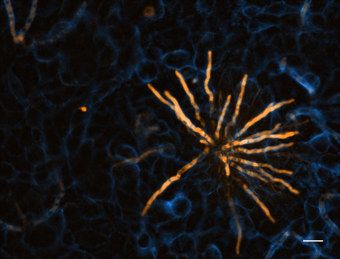
TdTomato-expressing Aspergillus fumigatus (amber) microcolony after 12 hours development on an A549 epithelial alveolar monolayer labelled with ConA-FITC (blue)
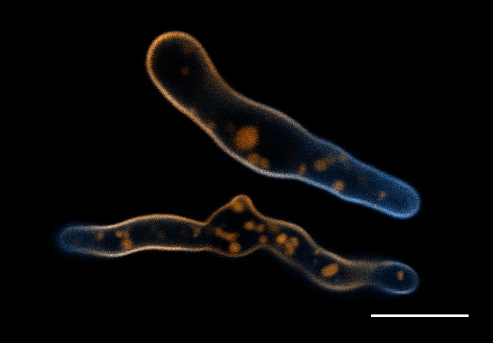
Aspergillus fumigatus germlings loaded with a synthetic cell membrane counterstain TMR-PAF96 (amber) and treated with fluorescent BODIPYcPAF26 anti-fungal peptide (cyan) to determine peptide’s localisation and mode of entry into the pathogen.
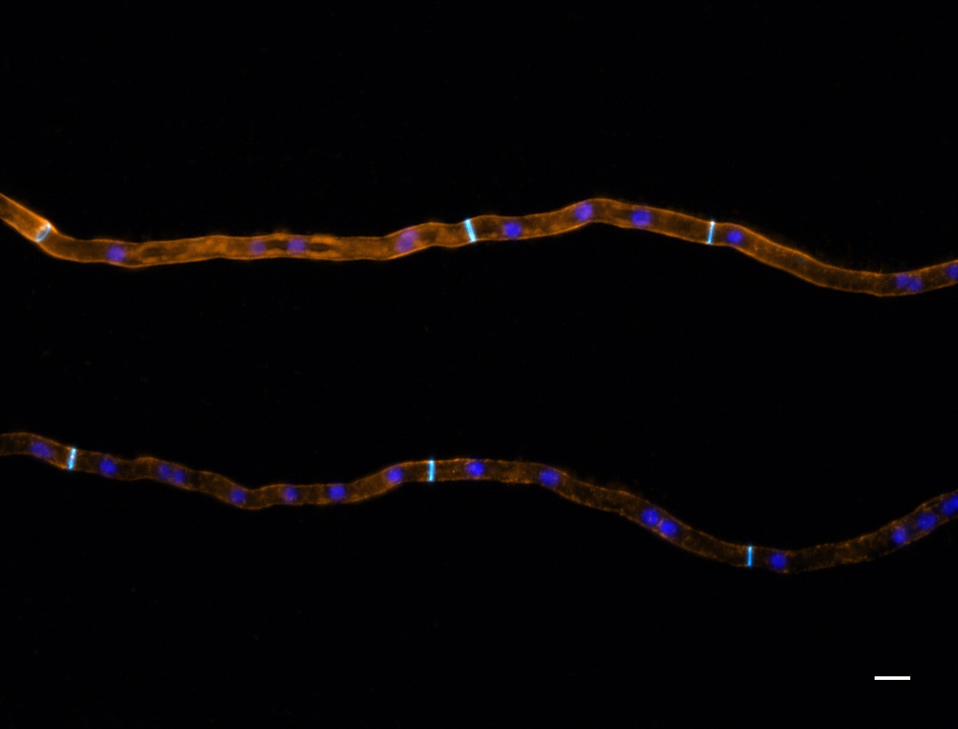
Neurospora crassa hyphal filaments treated with fluorescently labelled siRNA (amber) to determine its localisation, with respect to nuclei (blue) and the fungal cell wall and septa (cyan).
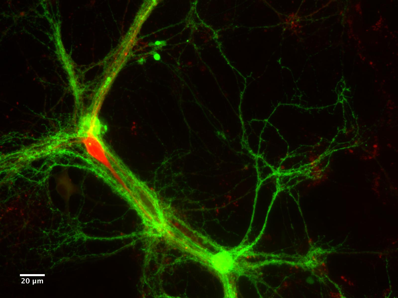
Ultrafast endocytosis at mouse hippocampal synapses taken with a CoolLED pE-2
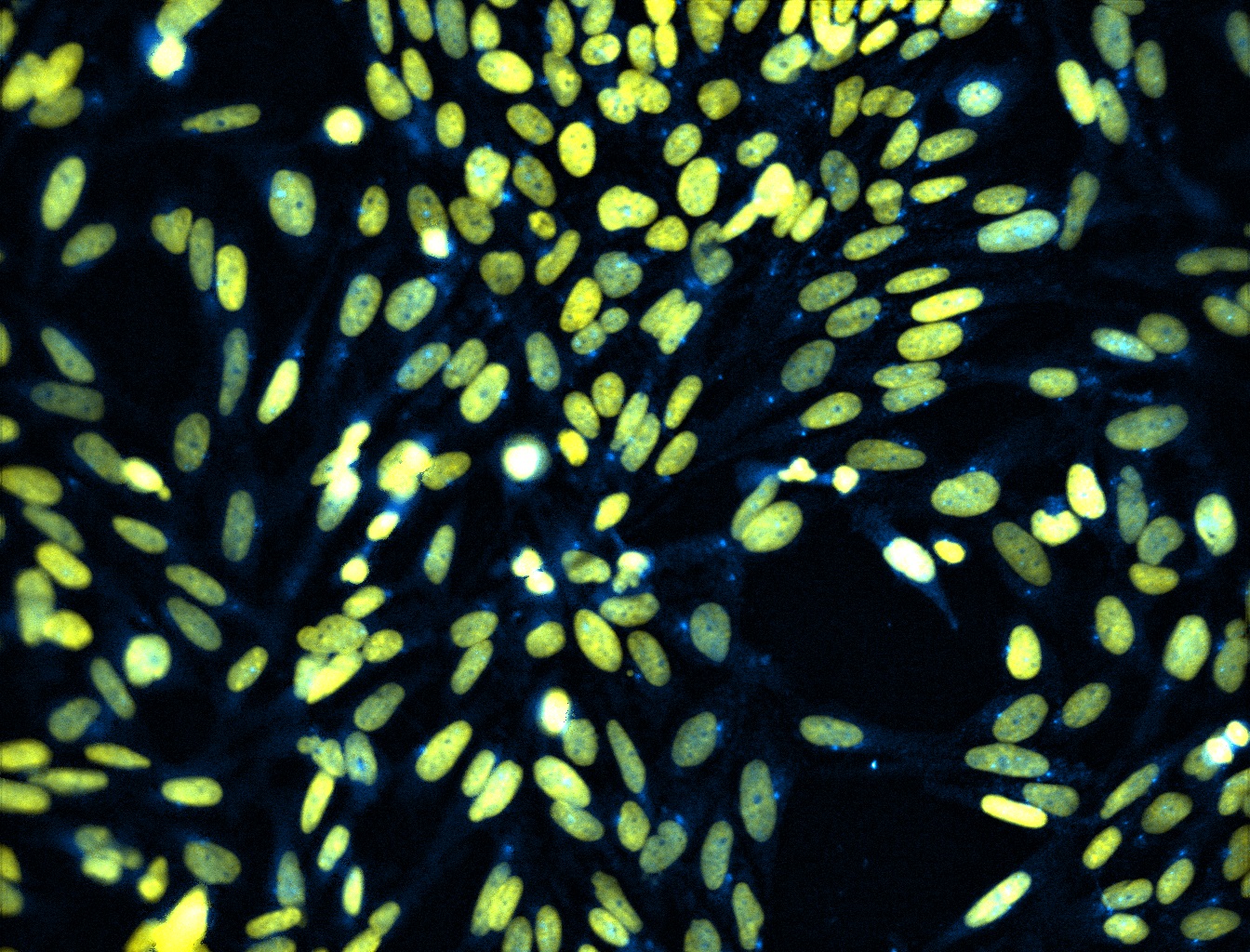
Antibody staining of a transcription factor localising in the nucleus
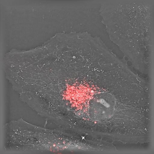
Live HeLa cells stained with lysosome marker imaged with Nanolive’s holotomographic technology.
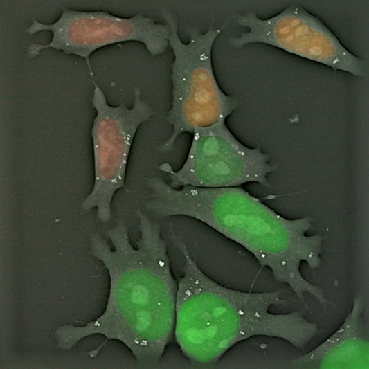
Live FUCCI mouse embryonic stem cells imaged with Nanolive’s holotomographic technology
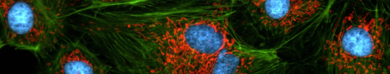
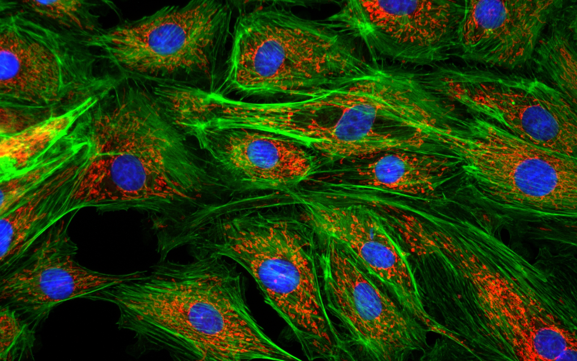
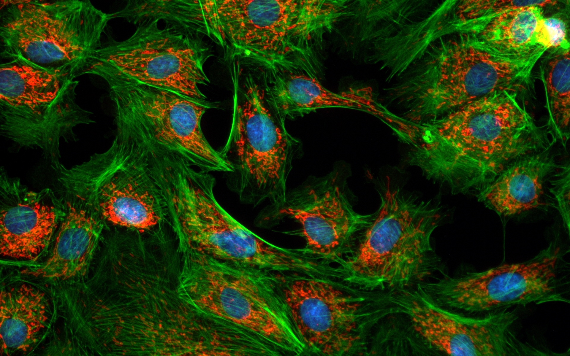
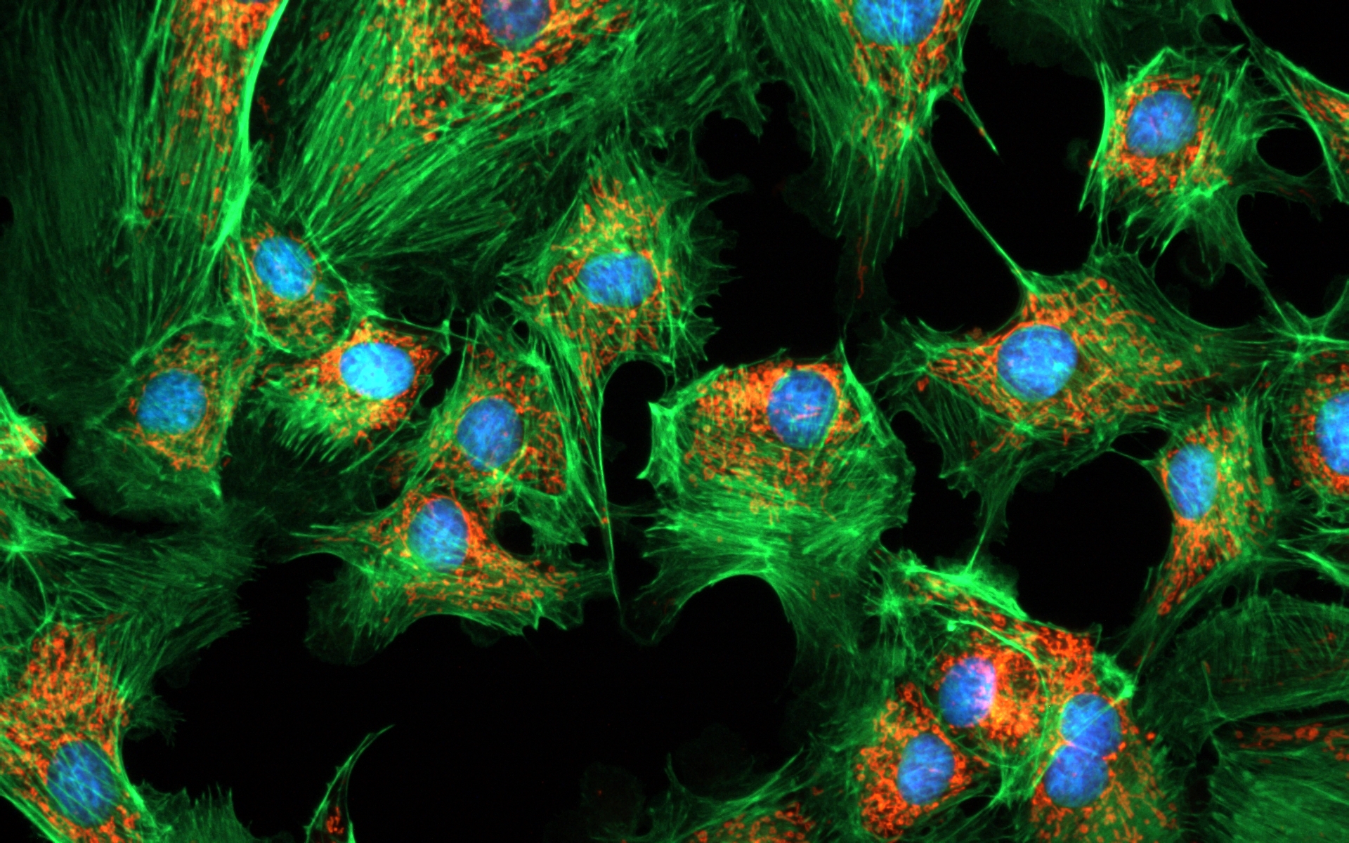
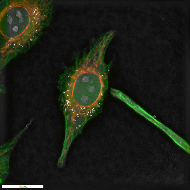
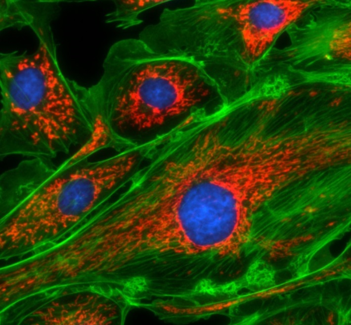
Fixed HeLa cells stained with Antibody for cytoskeleton (green), mitochondria (orange) and nuclear membrane(red). The whole cell was also imaged with Nanolive’s holotomographic technology and the four images were overlapped to create the overlay.
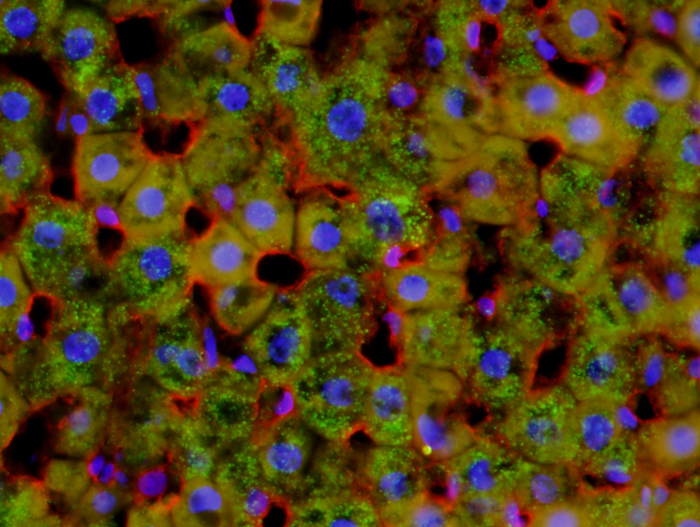
Fluorescent rat liver image taken using a CoolLED pE-300white, Olympus BX51 40x objective and a DP71 color camera
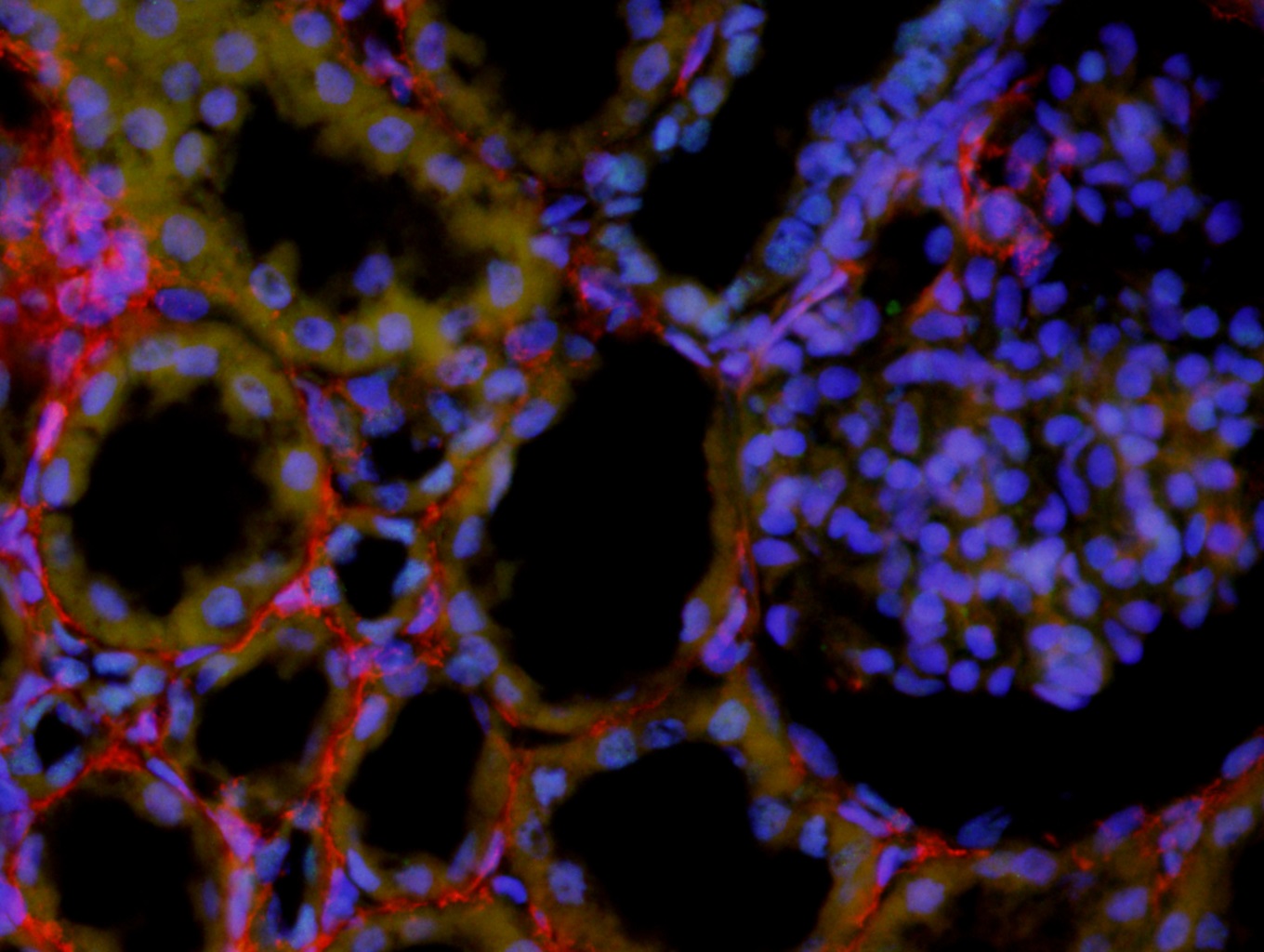
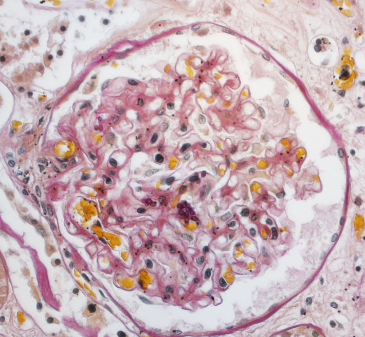
Fluorescent kidney image taken using a CoolLED pE-300white, Olympus BX51 40x objective and a DP71 color camera
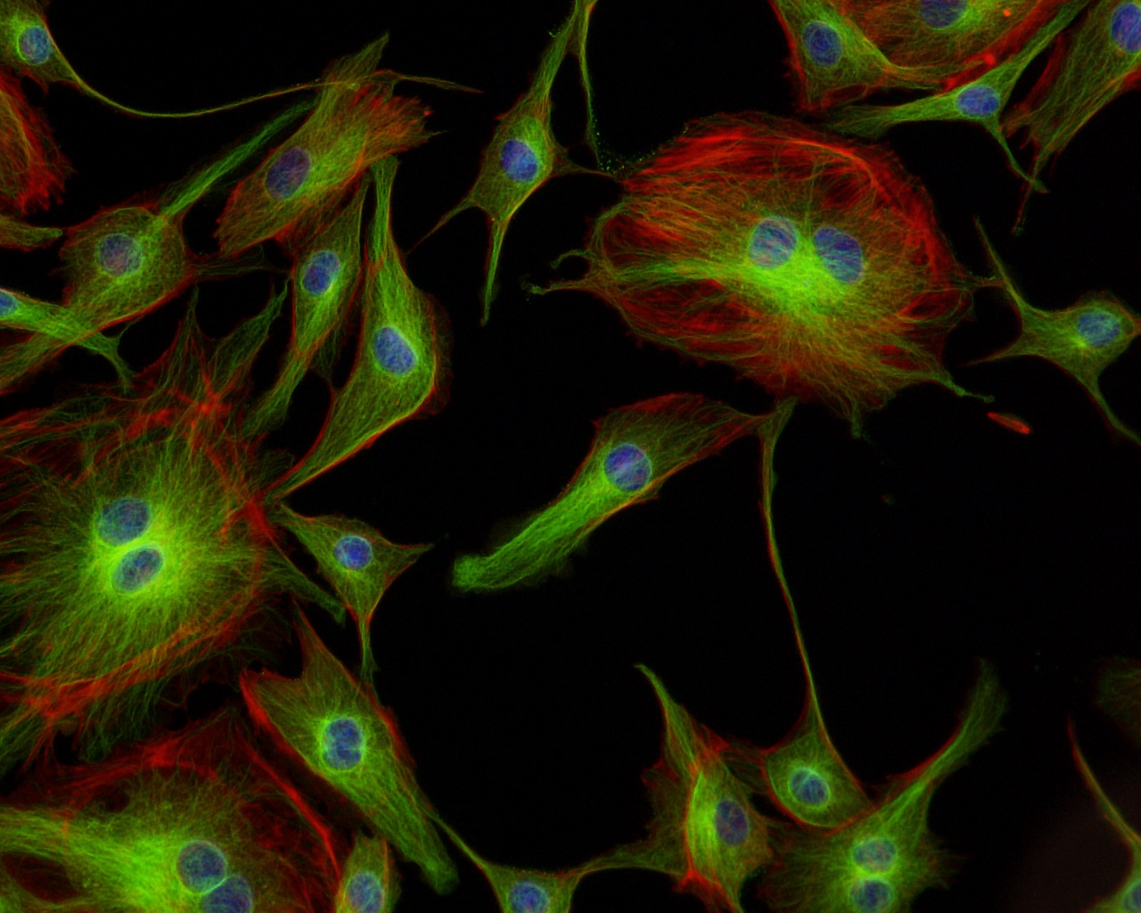
Fluorescent bovine pulmonary artery endothelial cells image taken using a CoolLED pE-300white, Nikon 20x objective and a black and white camera
Transmitted Podcarpus Elatus Leaf image taken using a CoolLED pE-100wht, Olympus BX51 20x objective and a DP71 color camera
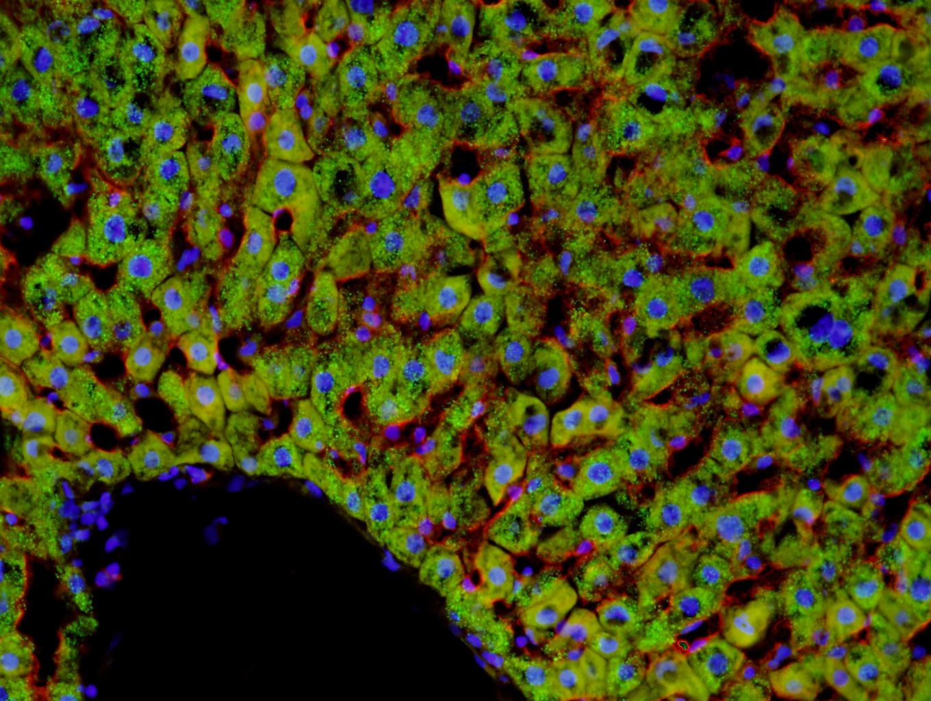
Fluorescent rat liver image taken using a CoolLED pE-300white, Olympus BX51 20x objective and a DP71 color camera
Fluorescent skin image taken using a CoolLED pE-300white, Olympus BX51 40x objective and a DP71 color camera
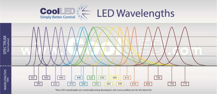
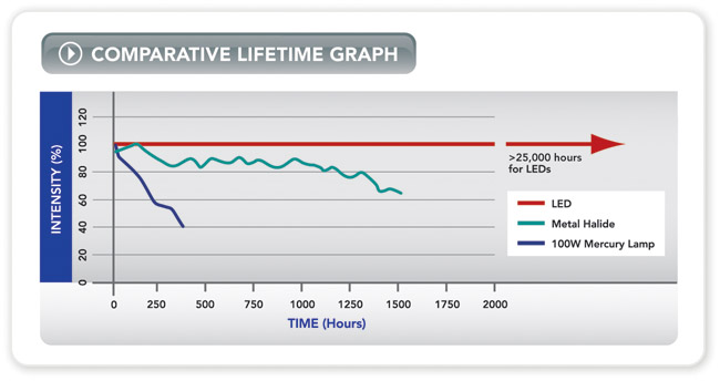
Broadest LED spectrum available: 365-770nm – replaces metal halide or mercury
Compatible with existing single and multi-band filter sets – no need to buy new filters
Instant on/off – no shutters required, no warm up or cool down
Simple to fit, simple to use – no alignment, a once only adjustment
Stable & repeatable – reliable and consistent results
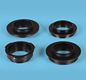
Wide range of microscope adaptors – fits most microscopes
Precise intensity control in 1% steps (0-100%) – no ND filters required
Excellent uniformity over field of view – fixed and stable, no alignment necessary
Long lifetime – expected to exceed 25,000 hours of operating time
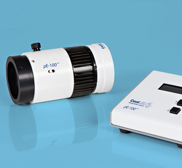
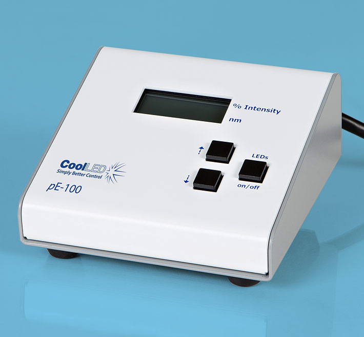
CoolLED pE-100
The pE-100 system has been developed for fluorescence applications requiring a single LED excitation wavelength. It is perfect for clinical applications such as routine screening (e.g. using Auramine for Tuberculosis), for research applications requiring precise intensity control and fast-switching, or for electrophysiology applications where light has to be delivered to a specific location. The user can select from 20 different LED wavelengths, ranging from the near-UV at 365nm to the near-IR at 770nm. The system comprises a pE-100 LED Light Source, control pod, and power supply.
There are three standard pE-100 configuration options:
Direct-fit
(pE-100) for connecting to a microscope (epi-port) by selecting from a range of microscope adaptors which covers all current and most older models. A simple once only adjustment will allow optimisation to the optical path of the microscope.
Liquid Light Guide
(pE-100light guide) with a fixed 3mm diameter, liquid light guideFiber
(pE-100fiber) with an SMA connection for accepting multimode fibers. The pE-100fiber has been designed with efficient coupling into a wide range of multimode fibers. The pE-100fiber also includes excitation filter holders.
Combining pE-100s:
Direct fit configuration
pE-100 units can be combined using the pE-Combiner for applications requiring a second LED wavelength with independent control and triggering.
Liquid light guide or multimode fiberusing a fixed two wavelength configuration can be specified. Two pE-100 Light Sources are combined, providing independent control and triggering. For further information go to the Downloads tab
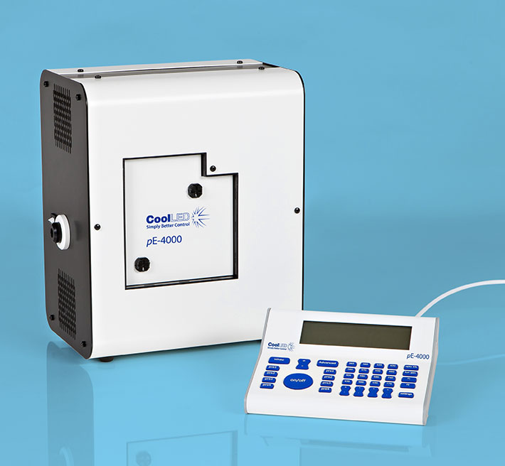
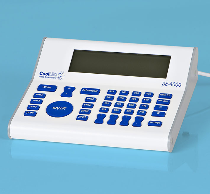
NEW GENERATION LEDs The pE-4000 sets the standard as the universal illumination system for fluorescence microscopy. The system has 16 selectable LED wavelengths across 4 channels that can be matched to the filters and fluorophores of almost any microscope, making it the broadest spectrum of illumination available.
The CoolLED pE-4000 now benefits from our award winning sustainable Greentechnology and delivers enhanced intensity at your microscope sample plane with a significant reduction in energy consumption, and is supplied with a 3 year warranty after registration.
The NEW Enhanced pE-4000 is a powerful 4-channel high-specification LED illumination system. It is flexible, controllable and environmentally friendly. Benefits include:
SELECTABILITY
Choice of 256 wavelength combinations from 16 installed LEDs (365nm -770nm)
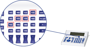
Choose 1 of 4 wavelengths for each 4 channels on the control pod
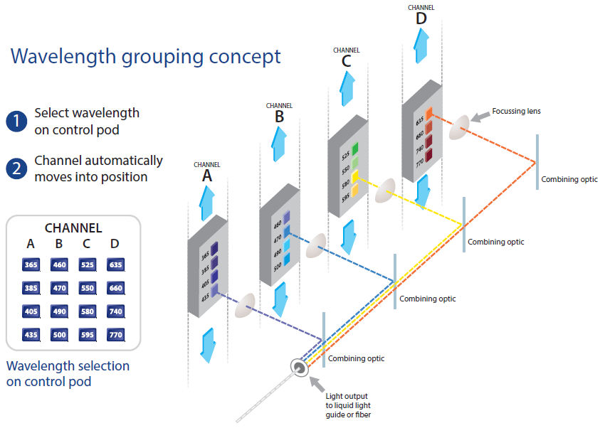
Unique wavelength grouping concept (see below)
Optimum wavelength to suit your experiment
Specific wavelength characterisation for optimal optogenetic control of cell responses
Wavelength Grouping Concept
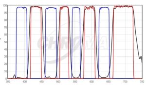
As the first company to introduce LEDs into fluorescence microscopy, CoolLED has developed a comprehensive understanding of the complexities of configuring and selecting filter sets for samples which have multiple stains. These ’multi-band’ filters must have a corresponding absorption and emission band for each stain and the bands must be kept spectrally separate to ensure there is no overlap.
DAPI/FITC/TRITC/Cy5 Quad filter excited by pE-4000 at matched wavelengths shown above.
CoolLED’s innovation comes from recognizing that all the stains used in multi-band work can have their absorption bands divided into four separate groups across the spectrum due to the restricted bandwidth available. This has allowed the development of a patent-pending, wavelength-grouping concept which makes it possible to deliver more power in an efficient four channel system.
With its unique 16 selectable LED wavelengths, the pE-4000 ensures optimum compatibility with all single and multi-band filter sets. The flexibility and extensive functionality of the pE-4000 broadens the range of illumination options in core facilities.
The pE-4000 allows multiple experiments with varying wavelengths on the same sample , as shown here.
(A) Representative photocurrent traces of a PhobosCA expressing CA1 cell evoked with different activation wavelengths and shutoff with 595nm light
(B) Photocurrent traces in the same cell evoked with 460nm light and shutoff with indicated wavelengths (10mW/mm2)
Wietek et al (2017) Anion-conducting channelrhodopsins with tuned spectra and modified kinetics engineered for optogenetic manipulation of behaviour, Scientific Reports volume 7, Article number 14957(2017) doi: 10.1038/s41598-017-14330-y
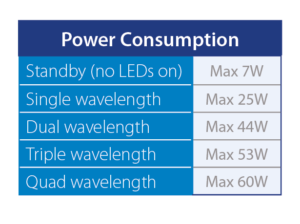
SUSTAINABILITY
Lowest Power Consumption – other LED technology uses 120-350 Watts
>25000 hours operation
Highly stable, repeatable LED technology
Market leading energy efficiency
CELL VIABILITY
Optimal illumination through control features to extend fluorescence of cells
Reduced photobleaching and phototoxicity
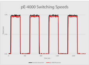
Microsecond switching
Variable pulse duration on/off
“When you can only control intensity of ‘white’ light (rather than individual channels), the level of photobleaching can be high. With the pE-4000, we can control the excitation of the individual channels. It is possible to optimise the excitation intensity according to the labelling, greatly reducing photobleaching and phototoxicity in a live experiment.” Dr Yan Gu, University of Sussex
“Suddenly, we were able to offer users uninterrupted extended live cell experiments of 100+ hours, without worrying about brightness fluctuations, lamps burning, room heating, etc. Also, users have reported markedly reduced bleaching and phototoxicity in their samples, both from the prokaryotic and the eukaryotic research fields.” Dr Jens Eriksson, Oslo University Hospital
CONTROLLABILITY
Advance integration into major microscope software platforms
Save wavelength combinations and return to previous set-up
USB interface
High speed TTL Triggering
Analogue control – dynamic intensity sinusoidal control option
White light control
Expansion Box
Individual channel intensity control
Pre-determined intensity saving option
VISIBILITY
High signal to noise ratio which gives cleaner images and data
“Striking, bright fluorescence images with a strong signal to noise ratio even when using a low magnification objective”
Graham Wright, Institute of Medical Biology, A*STAR Singapore
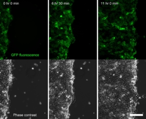
Time series of a confluent sheet of keratinocytes migrating during a live-cell imaging outgrowth assay, acquired on an Olympus IX-83 microscope equipped with a CoolLED pE-4000 lightsource (Fig. 4). Scale bar = 150 μm
ADAPTABILITY
Fits to all major research and clinical microscopes
Delivers light through a liquid light guide or fiber
Offers inline removable excitation filter holders
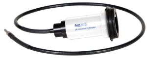
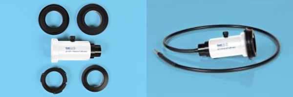
Uses the Universal Collimator
RELIABILITY
Assured through experience and QA assured processes
3 year Warranty after registration
First year Warranty swap
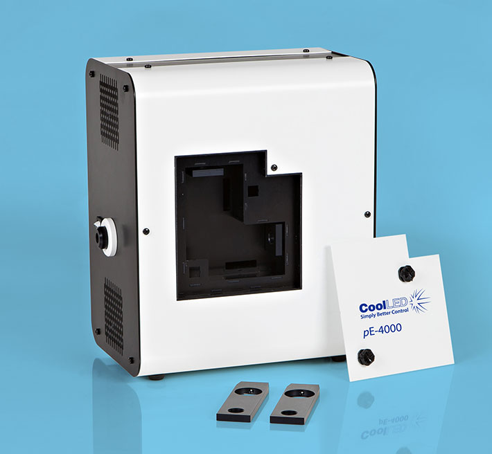
The compact pE-4000 Light Source houses all the LEDs, their combining optics, thermal management and control electronics. A dual mode control pod gives manual control in either White or Advanced modes. Light is delivered to the microscope by either a single liquid light guide (LLG) or fiber. LLGs can be used in conjunction with CoolLED’s pE-Universal Collimator which can accept a microscope adaptor from CoolLED’s extensive range.
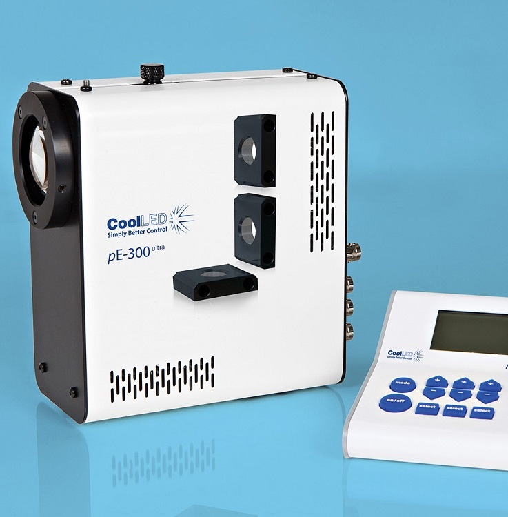
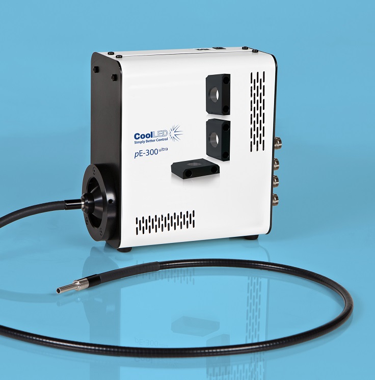
CoolLED’s pE-300ultra is a fluorescence microscopy light source which offers intense, broad-spectrum LED illumination for imaging most common fluorescent stains. Users have access to both microsecond switching via multiple TTL inputs and the ability to mount inline excitation filters. This, when paired with today’s high performance multi band filter sets, facilitates imaging traditionally done via a white light source and a filter wheel, with all the benefits of LED.
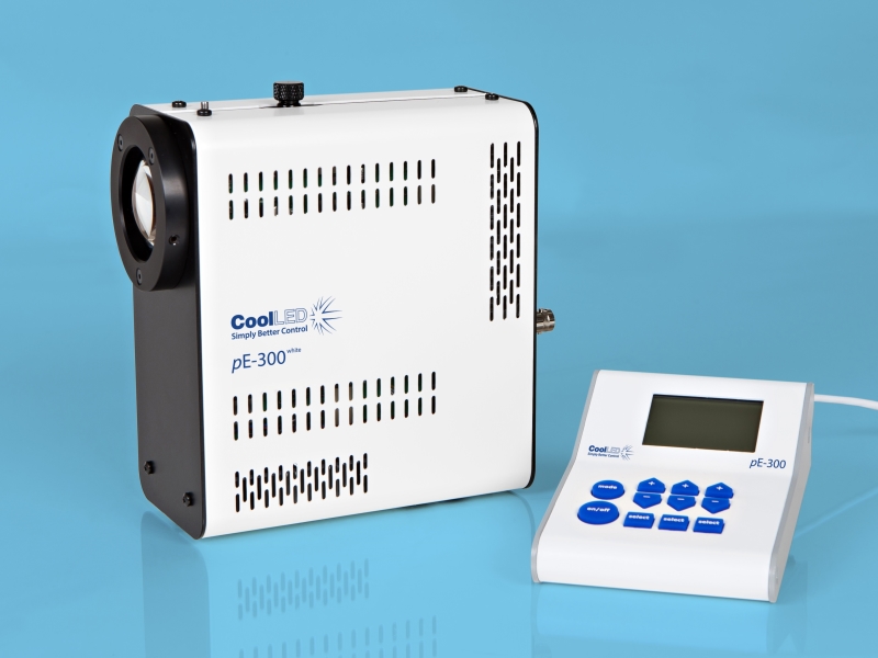
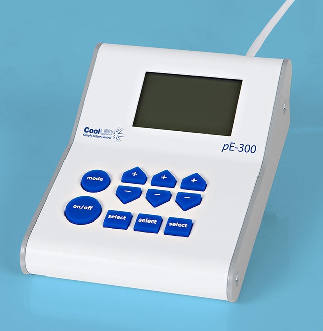
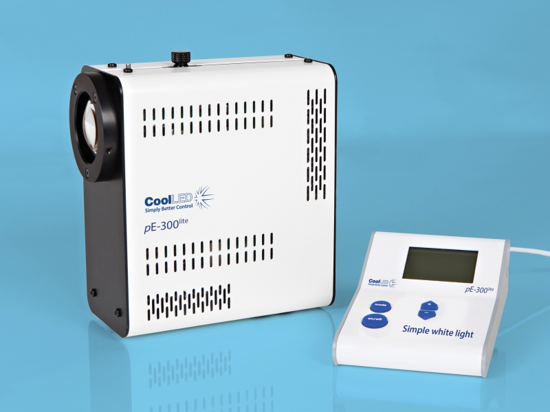
Control pod for CoolLED pE-300 ultra & white
Manual control for instant on/off
intensity control in 1% steps from 0 – 100%
The pE-300ultra is the most controllable member of the pE-300 Series. In addition to the many features of the pE-300lite and the pE-300white, the system offers precise control over wavelength intensity and shuttering. Until now these benefits have only been accessible to users of high end, highly flexible illuminators such as the CoolLED pE-4000.
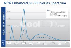
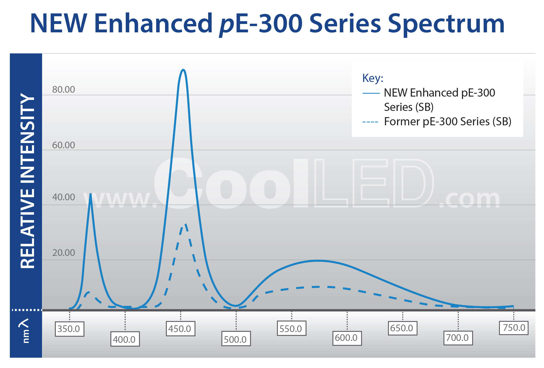
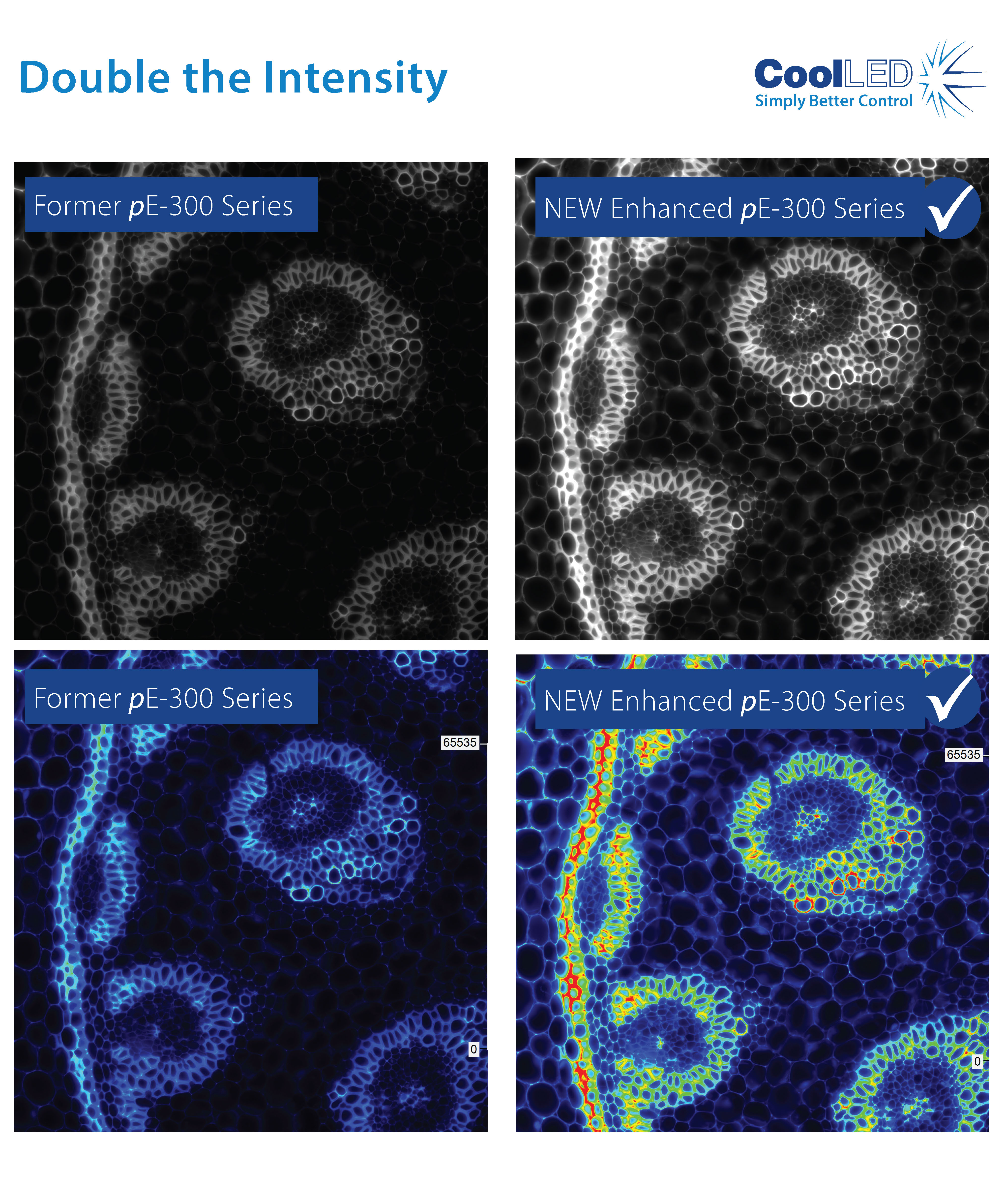
Throughout 2017 we will be releasing exciting new products and product updates. The first member of the CoolLED product range to receive the star treatment is the pE-300 Series.
As part of the award winning pE-300 Series, the NEW Enhanced pE-300ultra now delivers DOUBLE the intensity at the microscope sample plane. It allows adjustment of output in 1% steps, giving precise control. The broad spectrum covers everyday fluorophores: DAPI, CFP, Aqua, FITC, TRITC, TxRed, Cy5 and many more.
It is now possible to view and image your samples brighter than ever.
Convaleria taken with Photometrics Prime camera with 10ms exposure.
LED illumination means no mercury to dispose of and lower energy costs due to low power consumption and the precise control offered. With the new enhanced pE-300ultra, all 3 channels at full power use just 46 Watts. That is almost a quadruple efficiency increase!
This compares to other LED technology that uses 120 to 350 Watts. This leap forward in technology makes LEDs more attractive when compared to old Mercury or Metal Halide technology. Where “green” funding is available, the reduction in institutional energy usage improves the return on investment.
CoolLED’s pE-300 Series was the 2017 winner of the “Go Beyond Award” in the “Products” category for “Excellence in sustainability in laboratory and other high technology facilities” The International Institute for Sustainable Laboratories (I2SL) runs a unique awards program honouring organisations, individuals, products,and projects that are advancing sustainable, high-performance facilities.
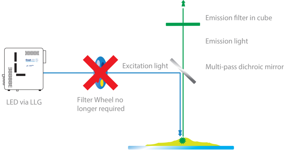
The pE-300ultra offers triggering from multiple TTL inputs which, coupled with the ability to mount inline excitation filters, provides microsecond switching of pre-filtered excitation light. This, when paired with today’s high performance multi-band filter sets, facilitates imaging traditionally done using a white light source and a filter wheel, with all the benefits of LED, and most excitingly at speeds not previously so affordable.
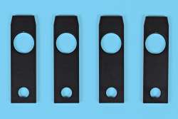
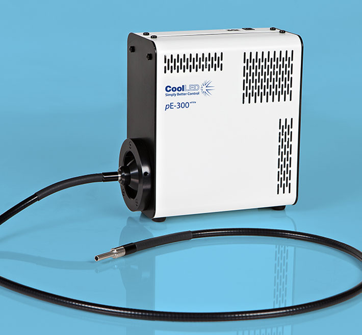
The system comprises a pE-300ultra Light Source, Control Pod, set of three Excitation Filter Holders and Power Supply. It can be specified with a microscope adaptor for direct fit configuration or with a 3mm diameter liquid light guide if there is a need to keep the source of illumination remote from the microscope. An optional pE-Universal Collimator and microscope adaptor can be selected for use with a liquid light guide.
A benefit of the pE-300ultra is that stains can be viewed either individually or in combination, without filter cube changes. This makes it ideal for use with multi-band filter sets as the screening process can be simplified when fewer filter cubes are used. Independent control of the three LED channels means that the user can control the level of excitation of each fluorescent stain independently on a multi-stained sample, potentially removing the need for single band filter sets altogether. For further information on improved multi-stain testing, go to the Downloads tab.
An important additional feature of the pE-300ultra is that it includes CoolLED’s “Sequence Runner” multiple channel excitation mode. Users can define the order of their fluorophore capture using their pE-300ultra Control Pod, then the pE-300ultra light source can accept a single TTL output from the experiment set-up’s camera to initiate the step-through of a sequence of excitation channels. This feature is independent of the individual channel TTL inputs on the light source. This offers users the facility to run through a sequence of excitation channels using a camera which has only a single TTL-out.
With an expected lifetime in excess of 25,000 on hours and a comprehensive range of microscope adaptors, the pE-300ultra can be fitted to most current and older microscopes and operate for many years without aligning or replacing bulbs. The result is a safe, convenient illumination system without any additional operating costs.
There are two pE-300ultra configuration options:
Direct-fit for connecting to a microscopes – by selecting from a range of microscope adaptors which covers all current and most older models. A simple once only adjustment will allow optimisation to the optical path of the microscope.
Liquid Light Guidewith a fixed 3mm diameter, liquid light guide. An optional pE-Universal Collimator can be specified in conjunction with a microscope adaptor if required.
The pE-300ultra offers:
Clean bright illumination across the spectrum – excites common fluorescent stains
Specify for existing single and multi-band filter sets – no need to buy new filters
Instant on/off – No shutters required, no warm up or cool down
Simple to fit, simple to use – no alignment, a once only adjustment
Stable & repeatable – reliable and consistent results
Wide range of microscope adaptors – fits most microscopes
Precise intensity control in 1% steps (0-100%) – no ND filters required
Excellent uniformity over field of view – fixed and stable, no alignment necessary
Long lifetime – expected to exceed 25,000 hours of operating time
Removable inline excitation filter holders – no moving parts
Individual channel triggering via TTL in microseconds
Sequence Runner – sequenced excitation from a single TTL-out
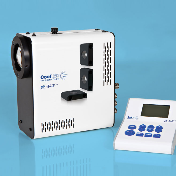
Utilising the successful pE-300 Series platform, the pE-340fura is a bespoke LED Illuminator for Fura-2 ratiometric calcium imaging, which also supports everyday fluorescence microscopy in a compact and affordable package.
The 340nm and 380nm LED illumination system provides the optimum excitation wavelengths for Fura-2-based calcium imaging allowing high-precision, stable, high-throughput imaging with video-rate time resolution.
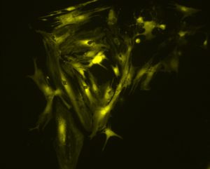
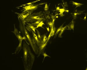
340nm Excitation
380nm Excitation
The images above show a field of cardiac myocytes (heart cells). The cells were loaded with Fura-2 using standard conditions (i.e. incubation with 2 micromolar Fura-2 acetoxymethyl ester for 30 minutes, followed by an additional 30 minutes for de-esterification.
Images were obtained by Martin Bootman and Katja Rietdorf, School of Life, Health and Chemical Sciences, The Open University, UK.
Until recently, the response time of illumination systems for Fura-2 imaging have been limited to milliseconds due to mechanical switching of the wavelengths in arc lamp and monochromator systems. However, the new pE-340fura can be controlled via convenient BNC TTL connections for precise illumination control in as little as 20 microseconds.
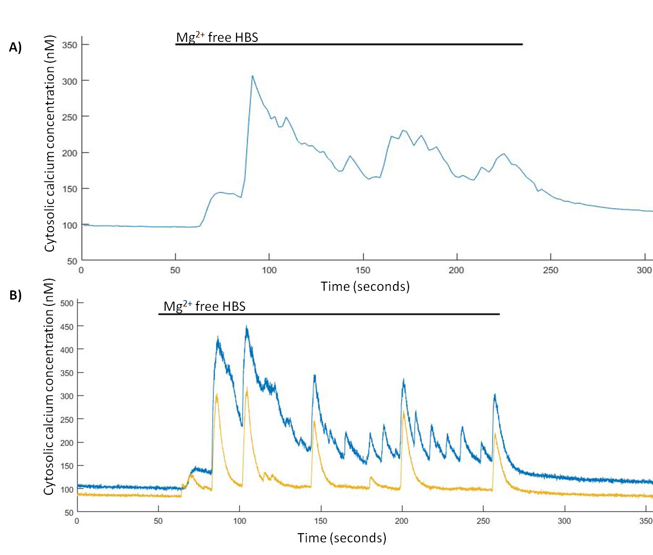
Spontaneous Ca2+ events are induced in Mg2+-free HBS. (A) Representative trace from a single hippocampal neuron of Mg2+-free induced Ca2+ events imaged at 0.5 Hz and (B) representative trace from two hippocampal neurons of Mg2+-free induced Ca2+ events imaged at 24.39 Hz.
TINNING, P. W., FRANSSEN, A. J. P.M., HRIDI, S. U., BUSHELL, T. J. and MCCONNELL, G. (2017), A 340/380 nm light-emitting diode illuminator for Fura-2 AM ratiometric Ca2+ imaging of live cells with better than 5 nM precision. Journal of Microscopy. doi:10.1111/jmi.12616
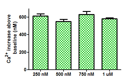
Using the new pE-340fura LED Illumination System, less Fura-2 dye can be loaded into the cells whilst still maintaining the same measured calcium concentration and good signal-to-noise ratio. The reduction in required dye not only improves cell-viability due to reduced dye toxicity, but also results in a cost reduction per experiment.
Comparison of Ca2+ increases obtained from the application of trypsin (100 nM) to tsA-201 cells loaded with different concentrations of Fura-2 AM.
TINNING, P. W., FRANSSEN, A. J. P.M., HRIDI, S. U., BUSHELL, T. J. and MCCONNELL, G. (2017), A 340/380 nm light-emitting diode illuminator for Fura-2 AM ratiometric Ca2+ imaging of live cells with better than 5 nM precision. Journal of Microscopy. doi:10.1111/jmi.12616
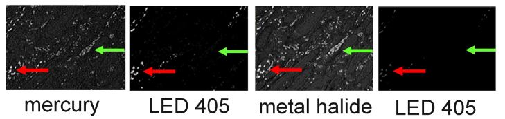
Work by Sandrine Prost et al., from the University of Edinburgh, has shown that with independent wavelength controllable LED sources, signal-to-noise is dramatically improved over bulb systems and even over some available white wide spectrum LED sources.
High levels of autofluorescence and fast photobleaching of specific fluorescence when illuminating Qdots with Metal Halide.
Prost S et al (2016) Choice of Illumination System & Fluorophore for Multiplex Immunofluorescence on FFPE Tissue Sections. PLoS ONE 11(9): e0162419. doi:10.1371/journal.pone.0162419
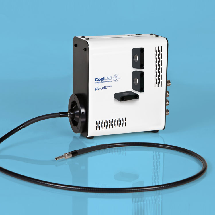
There are two pE-340fura configuration options:
Direct-fit for connecting to a microscopes – by selecting from a range of microscope adaptors which covers all current and most older models. A simple once only adjustment will allow optimisation to the optical path of the microscope.
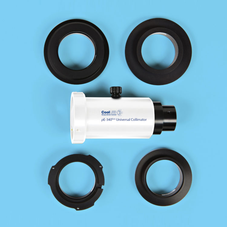
Liquid Light Guide with a fixed 3mm diameter, liquid light guide. An optional pE-340fura Universal Collimator can be specified in conjunction with a microscope adaptor if required, containing optics optimised to transmit the 340nm.