Advance Realtime Monitor. Easy-to-Use, Image Enhancement, etc
1. Information
ARM is image analysis software for JNOPTIC AcquCAM cameras. This S/W is interchangeable with all WDM cameras regardless of camera brand and model and even more the most strength point is simple and easy use of length, area, angle, etc.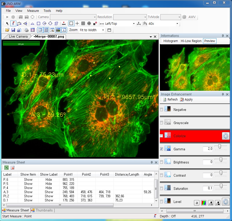
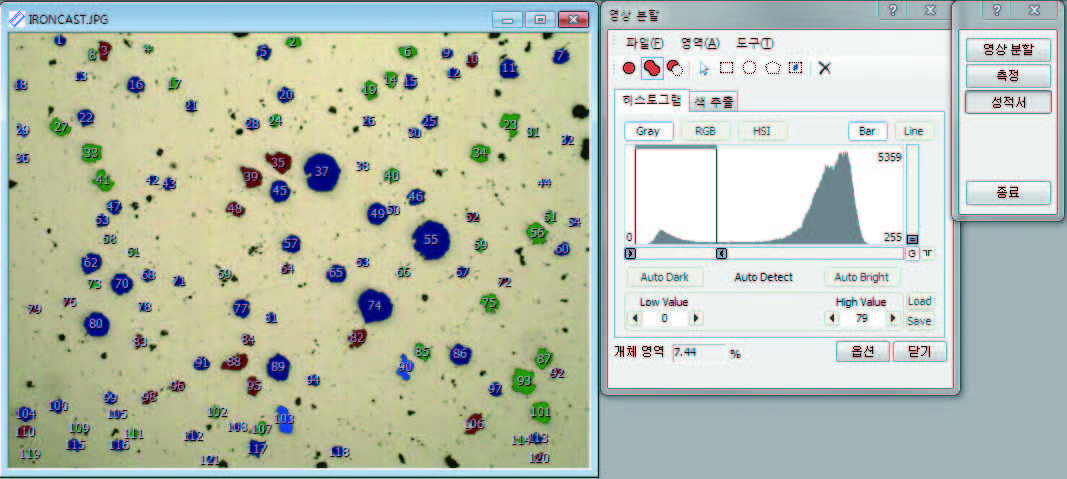
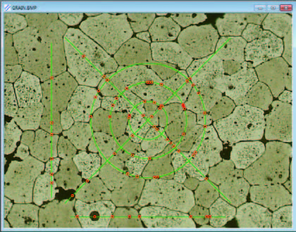
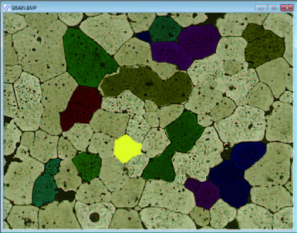
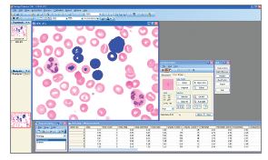
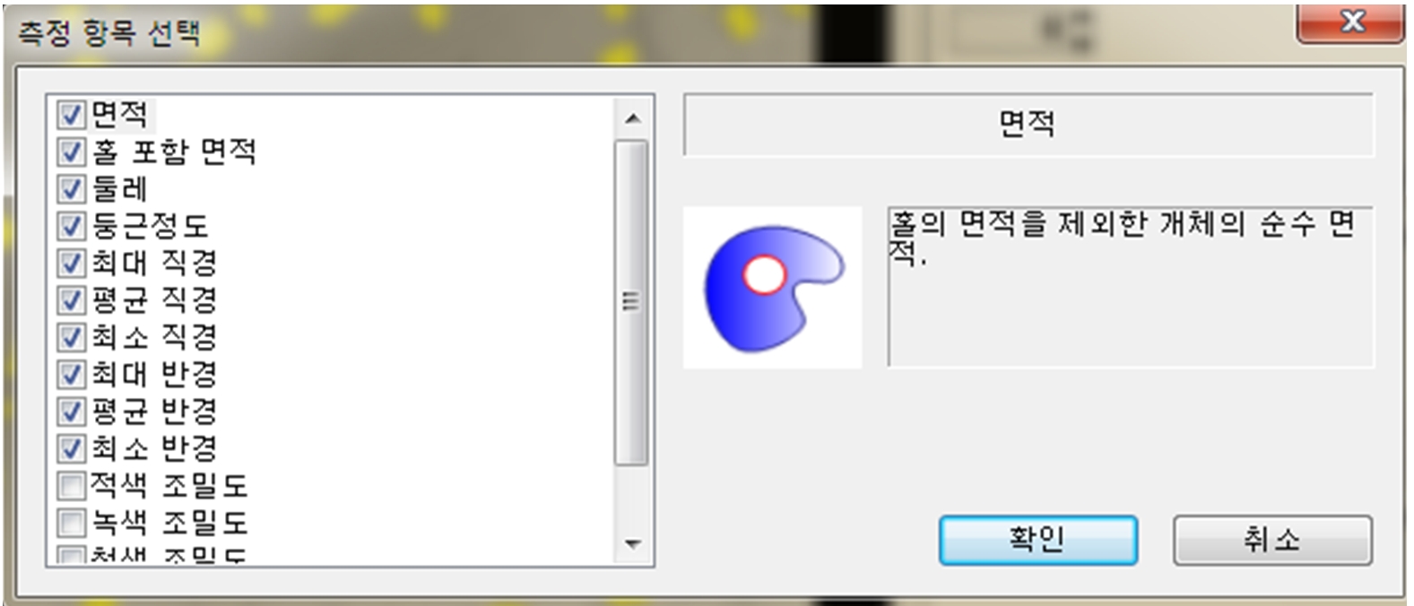
2. Simple measurement tools
![]()
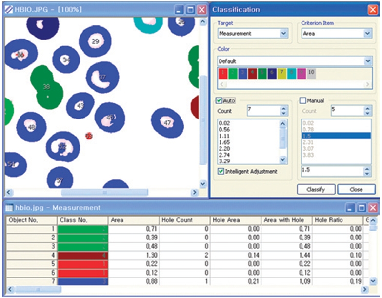
- Measurement Tools: Count, Distance, Angle, Area, etc.
- Guide Lines: Cross Line, Rectangular Lattice, Circular Grid
- Scale Panel on Live/Image Screen
- Available measurement on LIVE image and saved image mode
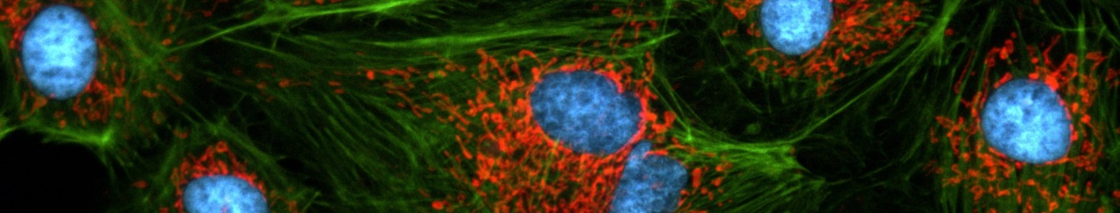
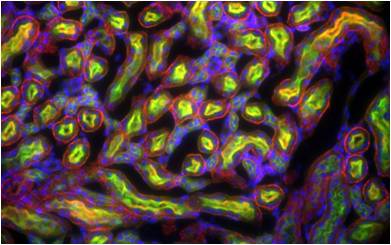
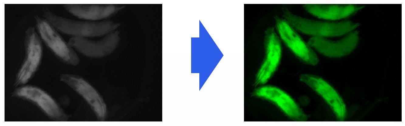
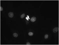
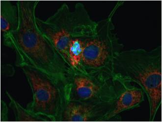
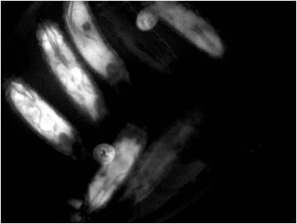
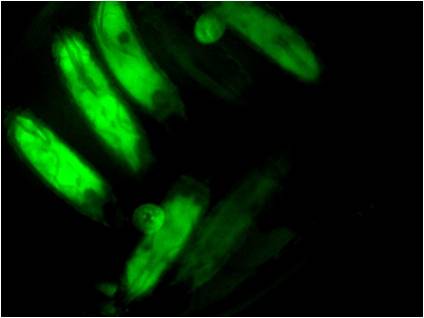
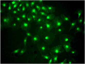
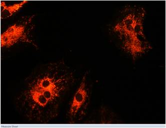
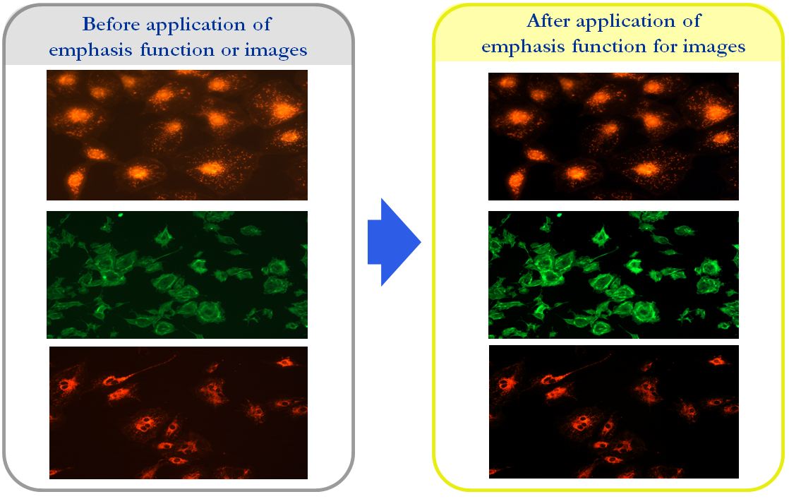
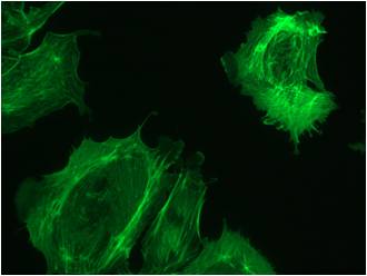
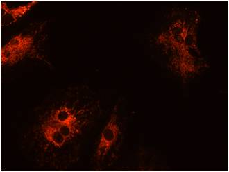
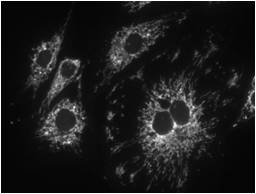
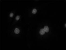
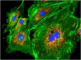
3-1. Specialization for observation of fluorescence images
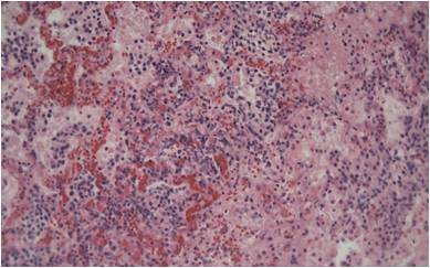
Improvement Function about Fluorescence Microscopy Image. (Used function: Auto Level, Low level, Pseudo Color, Merge Image)
Bottom on the left is merged image without improvement of image. And the bottom on the right is merged image after improvement of image. These images are shot by JNOPTIC AcquCAM Pro/G3 camera
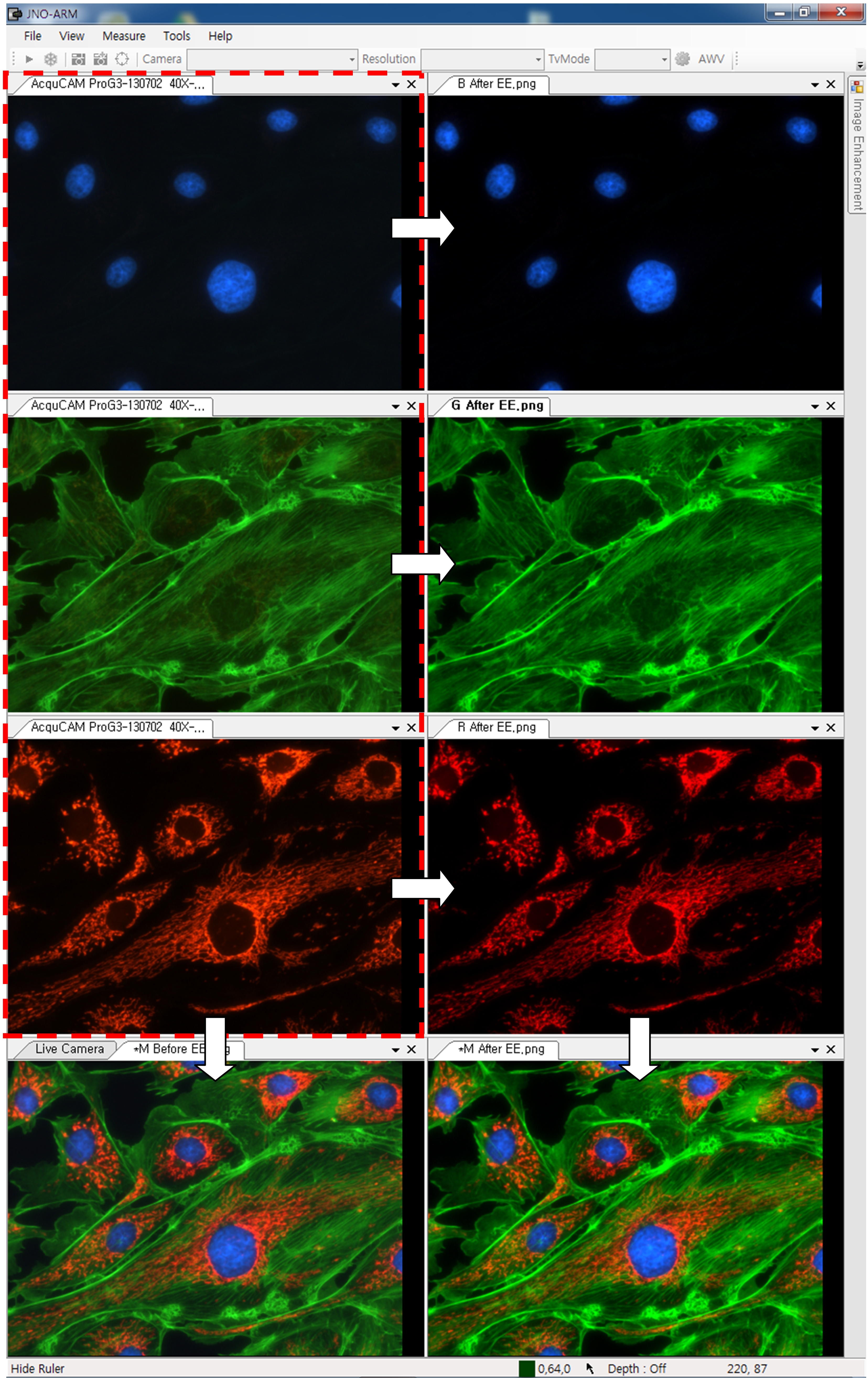
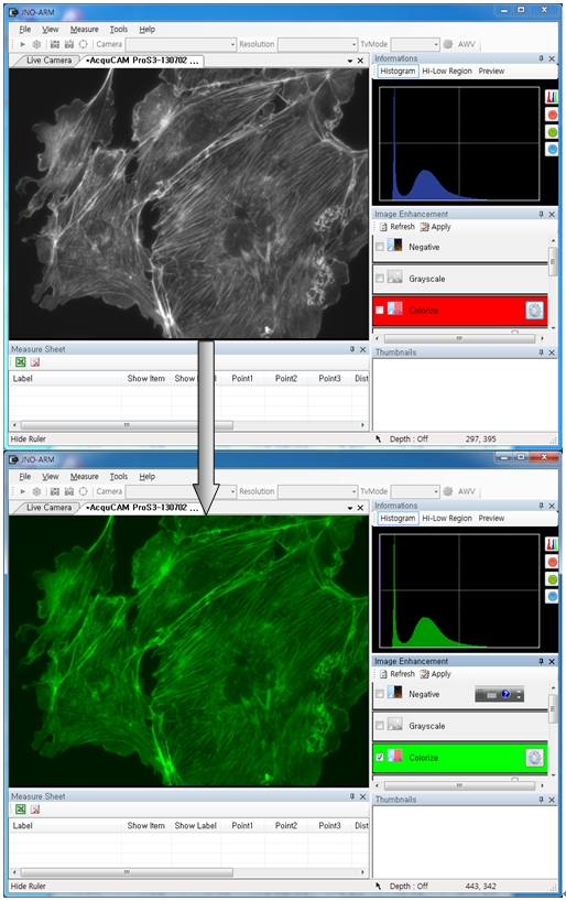
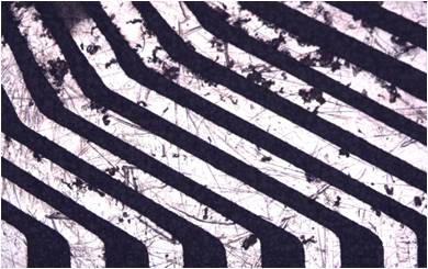
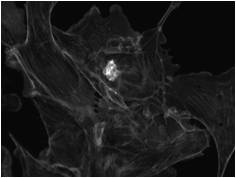
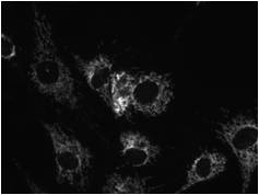
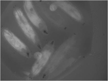
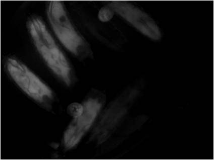
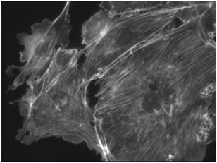
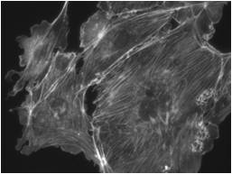
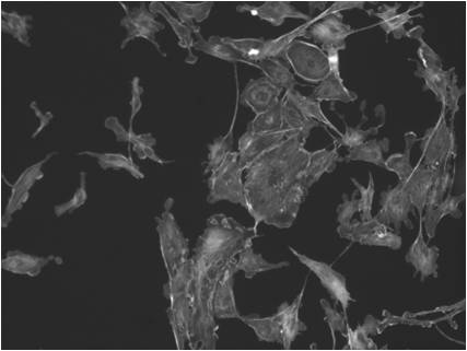
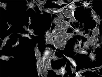
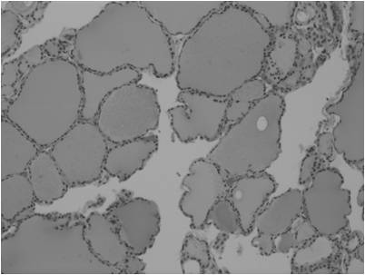
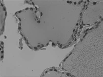
3-2-1 Effect of high-contrast image with simple operation (Monochrome)
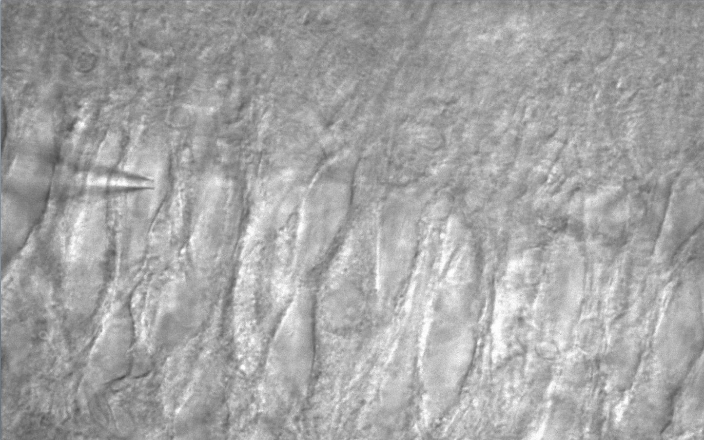
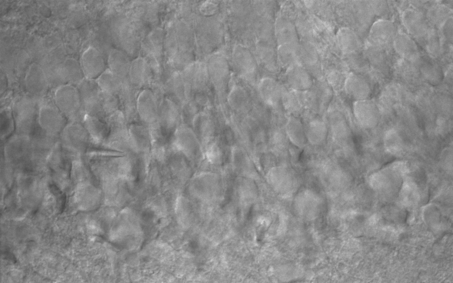
Improvement Function about Microscopy Image Monochrome. (Used function: Auto Level) Effective improvement of image with simple operation. This image is shot by JNOPTIC Pro/S3 camera
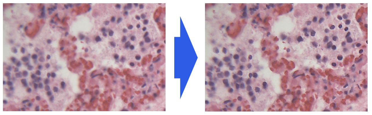
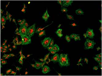
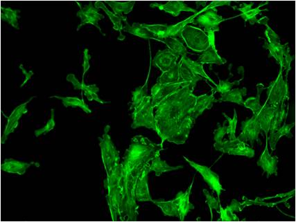
3-2-2 Effect of high-contrast image with simple operation (Color)
Improvement Function about Microscopy Image. (Used function: Auto Level)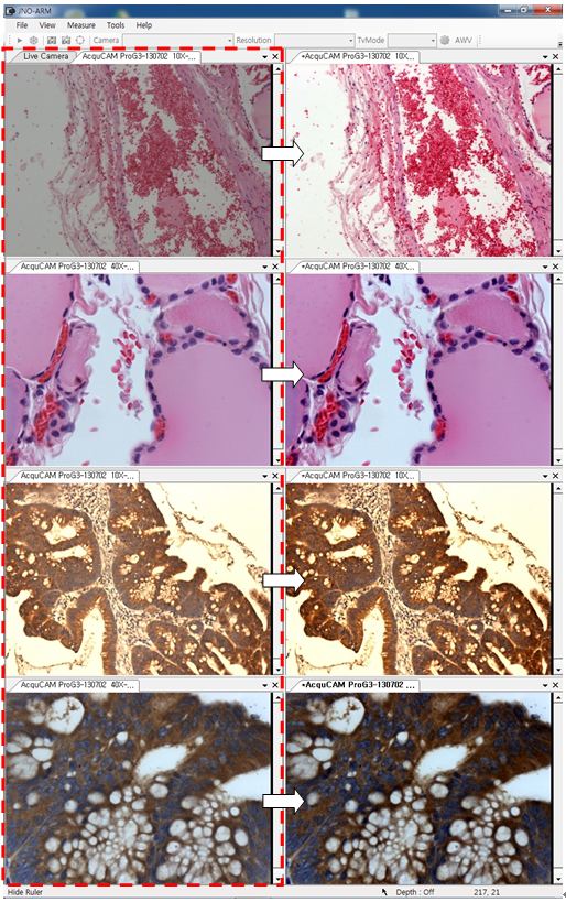 Effective improvement of image with simple operation. These images are shot by JNOPTIC AcquCAM Pro/G3 camera
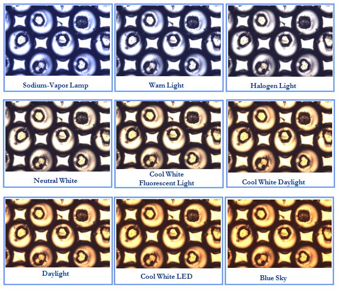
3-3-1 Live Pseudo Color (for color camera)
When you get fluorescence images using color camera, if the unwanted channel was shown because of cross talk, you could remove this channel to take advantage of function of Live Pseudo.
Improvement Function about Fluorescence Microscopy Live Camera Image. (Used function: Live Pseudo Color)
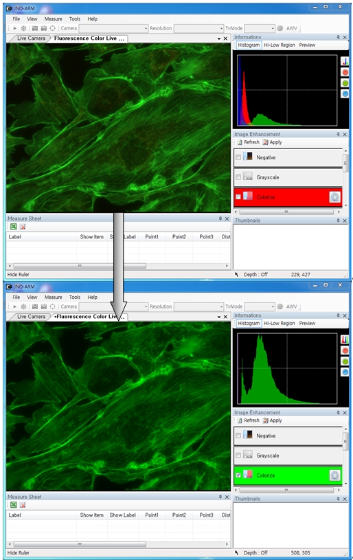
The extraction of wanted color information from color image (Remove BR on RGB source) These images are shot by JNOPTIC AcquCAM Pro/G3 camera
3-3-2 Live Pseudo Color (for Monochrome camera)
When you get fluorescence images using monochrome camera, you can raise efficiency of acquisition for fluorescence images to add similar false color to the color to be shown on the eyepiece in real time.
Improvement Function about Fluorescence Microscopy Live Monochrome Camera Image. (Used function: Live Pseudo Color)
Improvement of usage environment for monochrome camera to add false color on the image. These images are shot by JNOPTIC AcquCAM Pro/S3 camera
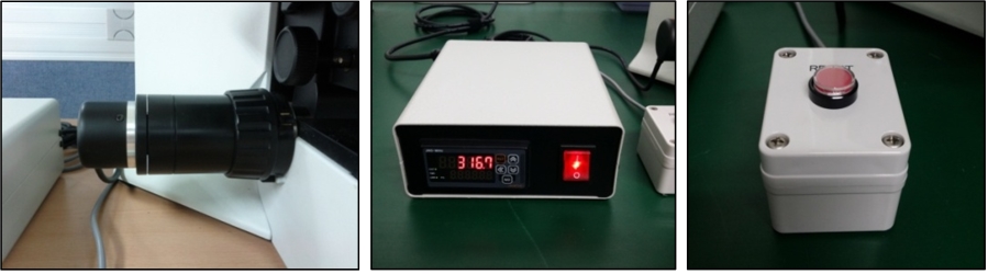
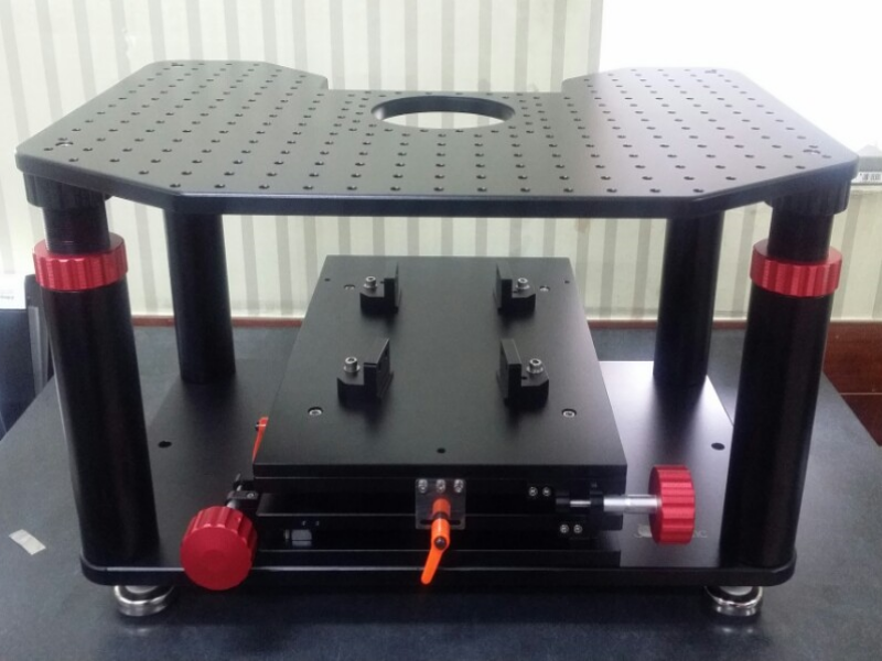
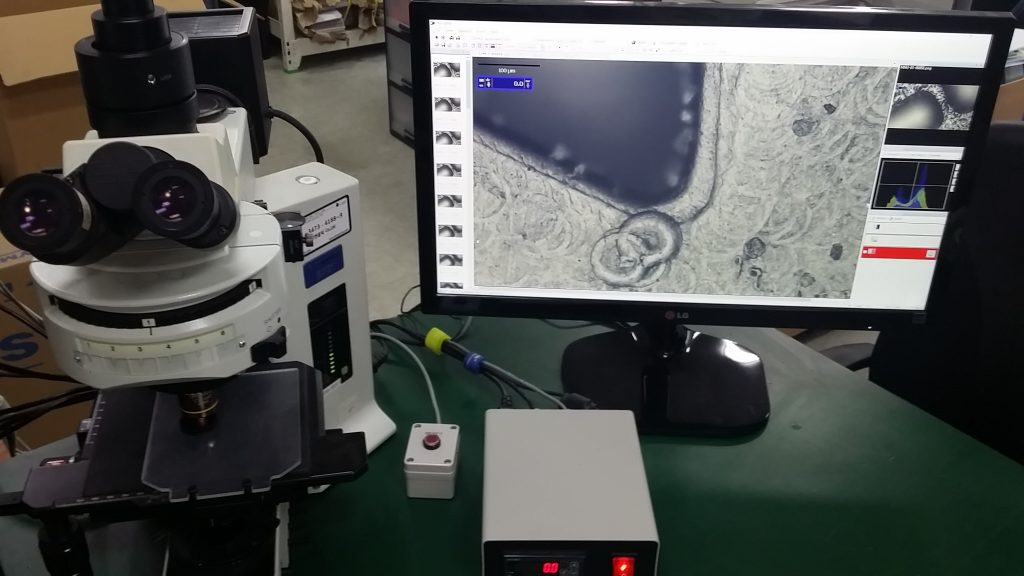
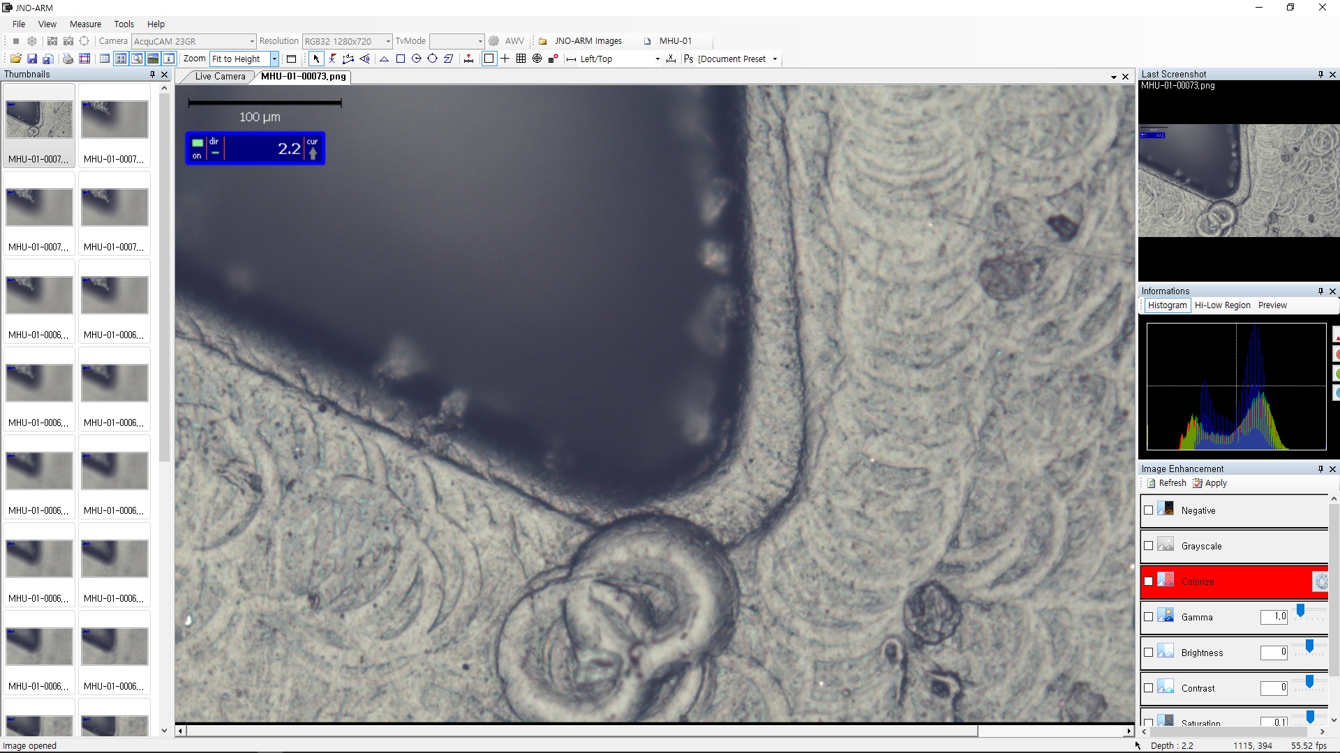
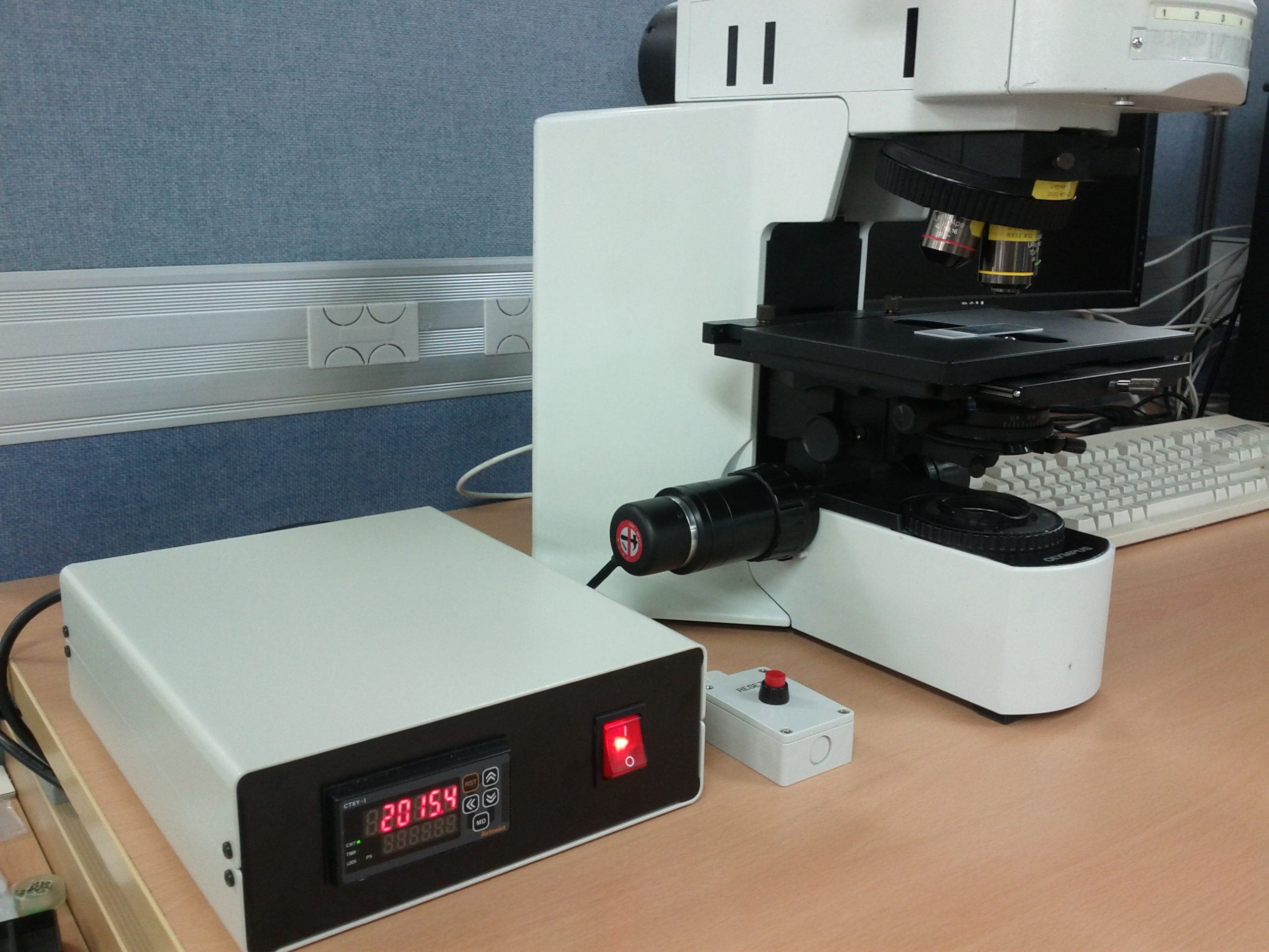
4. JNO-MHU(Option Unit)
Available measuring height with additional unit, JNO-MHU
※ JNO-MHU is sold separately as optional unit.
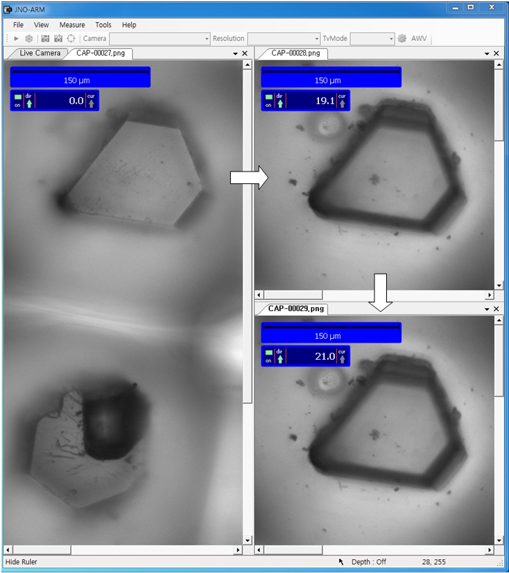
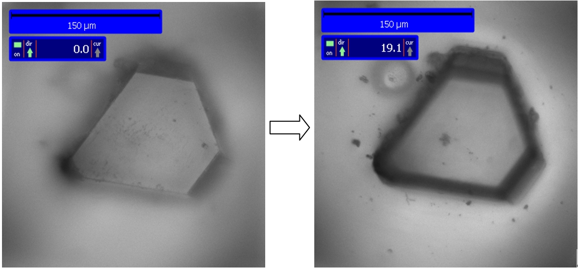
Left: Focus on top of sample (Z-axis reset), Right: Focus on bottom of sample (Z-axis height measurement)
< Measuring condition : over 20x objective, temp 20°C >
| Responding Model | Measurement value unit | Measurable height | Error of measuring 1000㎛ | Recommended measuring height |
| CX 41 | 0.2 ㎛ | -9.9 ~ 29.9㎜ | Below ±20㎛ | Below ± 2000㎛ |
| CKX 41 | 0.2 ㎛ | -9.9 ~ 29.9㎜ | Below ±20㎛ | Below ± 2000㎛ |
| BX – FM | 0.2 ㎛ | -9.9 ~ 29.9㎜ | Below ±20㎛ | Below ± 2000㎛ |
| BX 51/53 | 0.1 ㎛ | -9.9 ~ 29.9㎜ | Below ±10㎛ | Below ± 1000㎛ |
| MX 51 | 0.1 ㎛ | -9.9 ~ 29.9㎜ | Below ±10㎛ | Below ± 1000㎛ |
| MX 61L/61 | 0.1 ㎛ | -9.9 ~ 29.9㎜ | Below ±10㎛ | Below ± 1000㎛ |
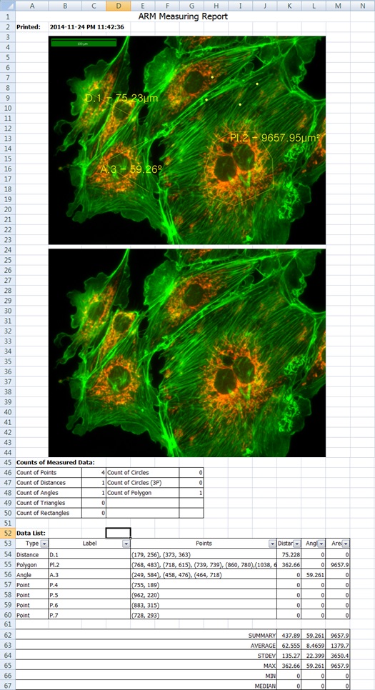
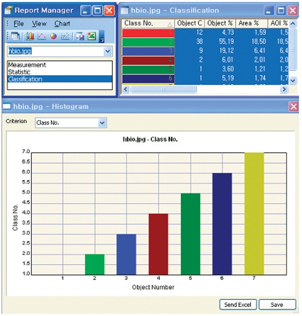
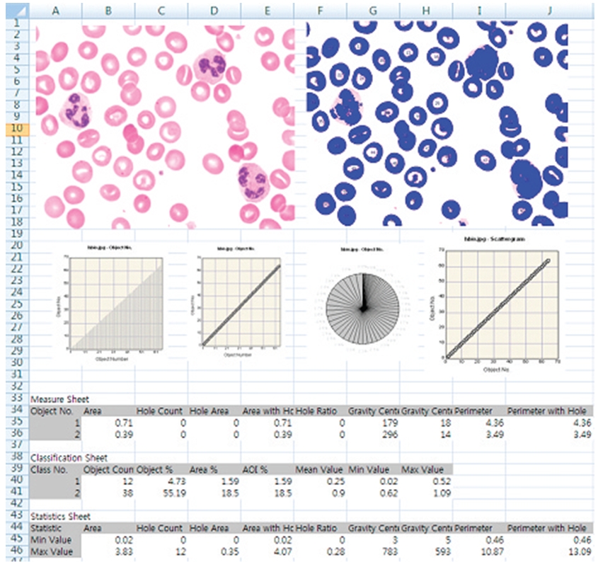
5. Reporting to Excel


