J.H.Jin의 모든 글

89402

JNOPTIC Filter 89402

ET – 391-32/479-33/554-24/638-31 Multi LED set

Quad band catalog set for Multi LED light engines like those manufactured by Lumencor, CoolLED, Excelitas and Cairn Research. Excitation passbands are matched to discrete, triggerable LEDs appropriate for excitation of DAPI/FITC/TRITC/Cy5 and similar fluors. Not intended for use with white light sources.

89402 Spectrum

Filters Type
89402x EX
89402bs BS
89402m EM


89402 Spec

NOTE: These require the use of filter wheels or sliders, or image-splitting devices to hold multiple individual filters.


69308

JNOPTIC Filter 69308

ET – ECFP/EYFP/mCherry with single band exciters

69308 Spectrum

Filters Type
ET430/24x EX
ET500/20x EX
ET577/25x EX
69008bs BS
69008m EM


69308 Spec

NOTE: sets with multi-band excitation filters are not appropriate for imaging with a monochrome camera.


69302

JNOPTIC Filter 69302

ET-DAPI/FITC/Texas Red® with single band exciters

69302 Spectrum

Filters Type
ET402/15x EX
ET490/20x EX
ET572/35x EX
69002bs BS
69002m EM


69302 Spec

NOTE: sets with multi-band excitation filters are not appropriate for imaging with a monochrome camera.


69008

JNOPTIC Filter 69008

ET-ECFP/EYFP/mCherry

This set is also ideal as a tripleband filter set in FISH applications for simultaneous viewing of commercially available Aqua, Green and Red FISH probes.

69008 Spectrum

Filters Type
69008x EX
69008bs BS
69008m EM

69008 Spec

NOTE: sets with multi-band excitation filters are not appropriate for imaging with a monochrome camera.


69015

JNOPTIC Filter 69015

ET-DAPI/Green/Red FISH

Triple band filter set ideal for simultaneous viewing of DAPI and commercially available Green and Red FISH probes. Attenuated level of DAPI excitaiton provides better balance of colors so that DAPI signal is not overwhelming.

69015 Spectrum

Filters Type
69015x EX
69015bs BS
69015m EM


69015 Spec

NOTE: sets with multi-band excitation filters are not appropriate for imaging with a monochrome camera.


69013v2

JNOPTIC Filter 69013v2

ET-DAPI/Green/Oragne #1 FISH

Triple band filter set ideal for simultaneous viewing of DAPI and commercially available Green and Orange FISH probes

69013v2 Spectrum

Filters Type
69013xV2 EX
69013bs BS
69013m EM


69013v2 Spec

NOTE: sets with multi-band excitation filters are not appropriate for imaging with a monochrome camera.


CoolLED Image Gallery

Sample : bovine pulmonary artery endothelial cell, BPAEC
CoolLED pE-340fura, OLYMPUS BX51, UPlanApo40x U-TV1XC
Filter Set : JNO-U(B), B(B), G(B)
Camera: JNOpTIC AcquCAM 23S2
상기 이미지는 각각의 B,G,R이미지를 JNO_ARM 소프트웨어로 머지한 이미지입니다.


Sample : bovine pulmonary artery endothelial cell, BPAEC
CoolLED pE-300ultra, OLYMPUS BX51, UPlanApo40x U-TV1XC
Filter Set : JNO-U(B), B(B), G(B),
Camera: JNOpTIC AcquCAM 23S2
상기 이미지는 각각의 B,G,R이미지를 JNO_ARM 소프트웨어로 머지한 이미지입니다.


Sample : bovine pulmonary artery endothelial cell, BPAEC
CoolLED pE-300ultra, OLYMPUS BX51, UPlanApo40x U-TV1XC
Filter Set : JNO-U(B), B(B), G(B),
Camera: JNOpTIC AcquCAM 23GR2
상기 이미지는 각각의 B,G,R이미지를 JNO_ARM 소프트웨어로 머지한 이미지입니다.


TdTomato-expressing Aspergillus fumigatus (amber) microcolony after 12 hours development on an A549 epithelial alveolar monolayer labelled with ConA-FITC (blue)


Aspergillus fumigatus germlings loaded with a synthetic cell membrane counterstain TMR-PAF96 (amber) and treated with fluorescent BODIPYcPAF26 anti-fungal peptide (cyan) to determine peptide’s localisation and mode of entry into the pathogen.


Neurospora crassa hyphal filaments treated with fluorescently labelled siRNA (amber) to determine its localisation, with respect to nuclei (blue) and the fungal cell wall and septa (cyan).


Ultrafast endocytosis at mouse hippocampal synapses taken with a CoolLED pE-2


Antibody staining of a transcription factor localising in the nucleus


Live HeLa cells stained with lysosome marker imaged with Nanolive’s holotomographic technology.


Live FUCCI mouse embryonic stem cells imaged with Nanolive’s holotomographic technology


Fixed HeLa cells stained with Antibody for cytoskeleton (green), mitochondria (orange) and nuclear membrane(red). The whole cell was also imaged with Nanolive’s holotomographic technology and the four images were overlapped to create the overlay.


Fluorescent rat liver image taken using a CoolLED pE-300white, Olympus BX51 40x objective and a DP71 color camera


Fluorescent kidney image taken using a CoolLED pE-300white, Olympus BX51 40x objective and a DP71 color camera


Fluorescent bovine pulmonary artery endothelial cells image taken using a CoolLED pE-300white, Nikon 20x objective and a black and white camera


Transmitted Podcarpus Elatus Leaf image taken using a CoolLED pE-100wht, Olympus BX51 20x objective and a DP71 color camera


Fluorescent rat liver image taken using a CoolLED pE-300white, Olympus BX51 20x objective and a DP71 color camera


Fluorescent skin image taken using a CoolLED pE-300white, Olympus BX51 40x objective and a DP71 color camera

CoolLED Control

pE-300 Series
CoolLED pE-300 Series Datasheet
CoolLED pE-300lite Exploded Diagram
CoolLED pE-300white Exploded Diagram
CoolLED pE-300ultra Exploded Diagram
CoolLED pE-300ultra Optogenetics Leaflet
CoolLED pE-300 Series User Manual 
CoolLED pE-300 Series Quick Start Guide
CoolLED pE-300 Series Troubleshooting Guide
CoolLED Driver File
CoolLED pE-300 Series Essential Commands Manual
CoolLED pE-Exerciser
CoolLED Firmware Update Program 
CoolLED pE-300 Series Firmware Update Instructions  

pE-340fura
CoolLED pE-340fura Datasheet
CoolLED pE-340fura Exploded Diagram
CoolLED pE-340fura User Manual
CoolLED pE-340 Quick Start Guide
CoolLED pE-340fura Troubleshooting Guide
CoolLED pE-340fura Essential Commands Manual 

pE-4000
CoolLED pE-4000 User Manual
CoolLED pE-4000 Quick Start Guide
CoolLED pE-4000 Troubleshooting Guide
CoolLED pE-4000 leaflet 
CoolLED pE-4000 Exploded Diagram
CoolLED Driver File 
CoolLED pE-4000 Essential Commands Manual 
CoolLED pE-Exerciser
CoolLED Firmware Update Program 
CoolLED pE-4000 Firmware Update Instructions

Remote via USB (B type) for imaging software control
Company Software Summary Screenshot Quick Start
Andor Andor iQ Click Here Click Here Click Here
Hamamatsu HCImage Click Here Click Here Click Here
LaVision BioTec ImSpector Pro Click Here Click Here
Leica LASX Click Here Click Here Click Here
Micromanager Micromanager Click Here Click Here Click Here
Molecular Devices MetaMorph Click Here Click Here Click Here
Nikon NIS Elements Click Here Click Here Click Here
Olympus CellSens Click Here Click Here Click Here
Visitron Systems Visiview Click Here Click Here
Intelligent Imaging Innovations (3i) Slidebook Click Here Click Here Click Here
Zeiss ZEN Click Here Click Here

VF-310-0Z Vibratome

Compresstome® VF 310-0Z Vibrating Microtome

CompresstomeⓇ VF-310-0Z 의 특징 : 

  • fresh 하고 고정된 tissue 모두 절단 가능
  • 절단 중 Z축으로의 이동을 제거하는 Auto Zero-Z 특허 기술
  • 일관되고 균일한 슬라이스 제작
  • 버퍼 트레이의 이동으로 간편한 세척이 가능
  • 블레이드 홀더의 탈착이 쉬움 (수동 얼라인먼트가 필요없음)
  • 직경 20mm 까지의 tissue  절단 가능
  • 사용하기 쉬운 컨트롤이 있는 단일 박스
  • 슬라이스 두께 : 4µm~ 1000µm ( 1µm 정밀도)
  • 슬라이스 넘버 카운터
  • 토탈 슬라이스 두께 카운터
  • 내구성을 위한 고품질의 디자인
  • 채터 마크 제거


VF-310-0Z 의 적용 가능 분야 :

  • 전기생리학 : brain slices, cardiac or myocardiac recordings, and kidney slices
  • 면역조직화학 : 4µm 만큼 얇은 고정 tissue 용
  • 기관형배양 : cultured slice experiments
  • 정밀절단 : lung, liver, kidney, spleen slices
  • 이미징 : slices 의 형광 이미지
  • 인시투하이브리디제이션 : RNA 검출
  • 전자현미경 : 마이크로아키텍쳐와 세포 해부학에 관한 연구

VF-310-0Z 의 Technical Spec :

 Technical Specification VF-310-0Z
Advance Speed 0-20 ㎜ / s, adjustable
Return Speed 20 ㎜/s
Vibration Frequency 0-45 Hz, adjustable
Vibration Amplitude 2 ㎜
Z-axis Vibration ~0 ㎛
Blade stainless steel, ceramic,             tungsten carbide,
sapphire diamond
Cutting Angle 15 degrees,
(no need for blade alignment)
Thickness Adjustment automatic or manual
Micrometer Resolution 1  ㎛
Maximum Tissue
Diameter
12.5 ㎜ (standard)
22㎜ (extra large)
15.5 ㎜ (large)
6.5㎜ (small)
Maximum Tissue
Length
30 ㎜
Minimum Slice
Thickness
4 ㎛
Cutting Mode single/ continuous
selectable
Bath 140 x 60 x 30 ㎜
Power Source 120V AC
Power Consumption 18 W
Dimension ( L x W x H ) 330 x 240 x 190 ㎜
Weight 5 ㎏

Down load  Technical Specification_VF-310-0Z

Training  Videos :

Using the Buffer Tray
Diagramming the Control Box
Attaching a New Blade
Plating Your Slices
Making the agarose solution for tissue embedding
Cleaning the specimen tube (part 1) for Compresstome® 
Cleaning the specimen tube (part 2) for Compresstome®
Transferring tissue slices cut with Compresstome®
Embedding tissue samples into specimen tubes
Starter Kit contents for the Compresstome®


VF-310-0Z 부품 : 


VF-310-0Z  소모품 : 


pE-100

The pE-100 offers:

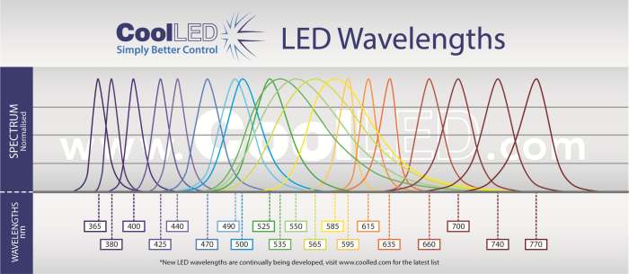
  • Broadest LED spectrum available: 365-770nm – replaces metal halide or mercury
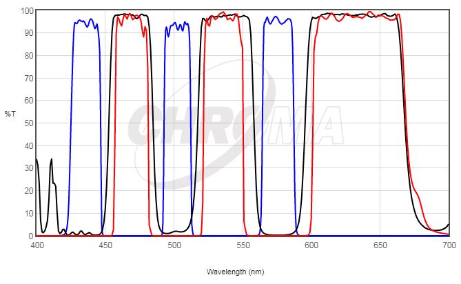
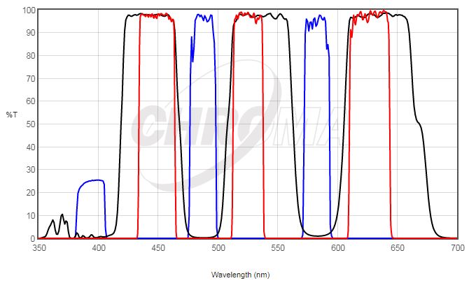
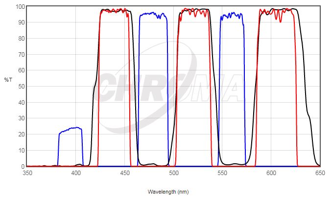
  • Compatible with existing single and multi-band filter sets – no need to buy new filters
  • Instant on/off  – no shutters required, no warm up or cool down
  • Simple to fit, simple to use – no alignment, a once only adjustment
  • Stable & repeatable – reliable and consistent results
  • Wide range of microscope adaptors – fits most microscopes
  • Precise intensity control in 1% steps (0-100%) – no ND filters required
  • Excellent uniformity over field of view – fixed and stable, no alignment necessary
  • Long lifetime – expected to exceed 25,000 hours of operating time

CoolLED pE-100


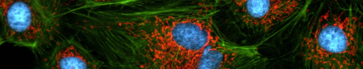
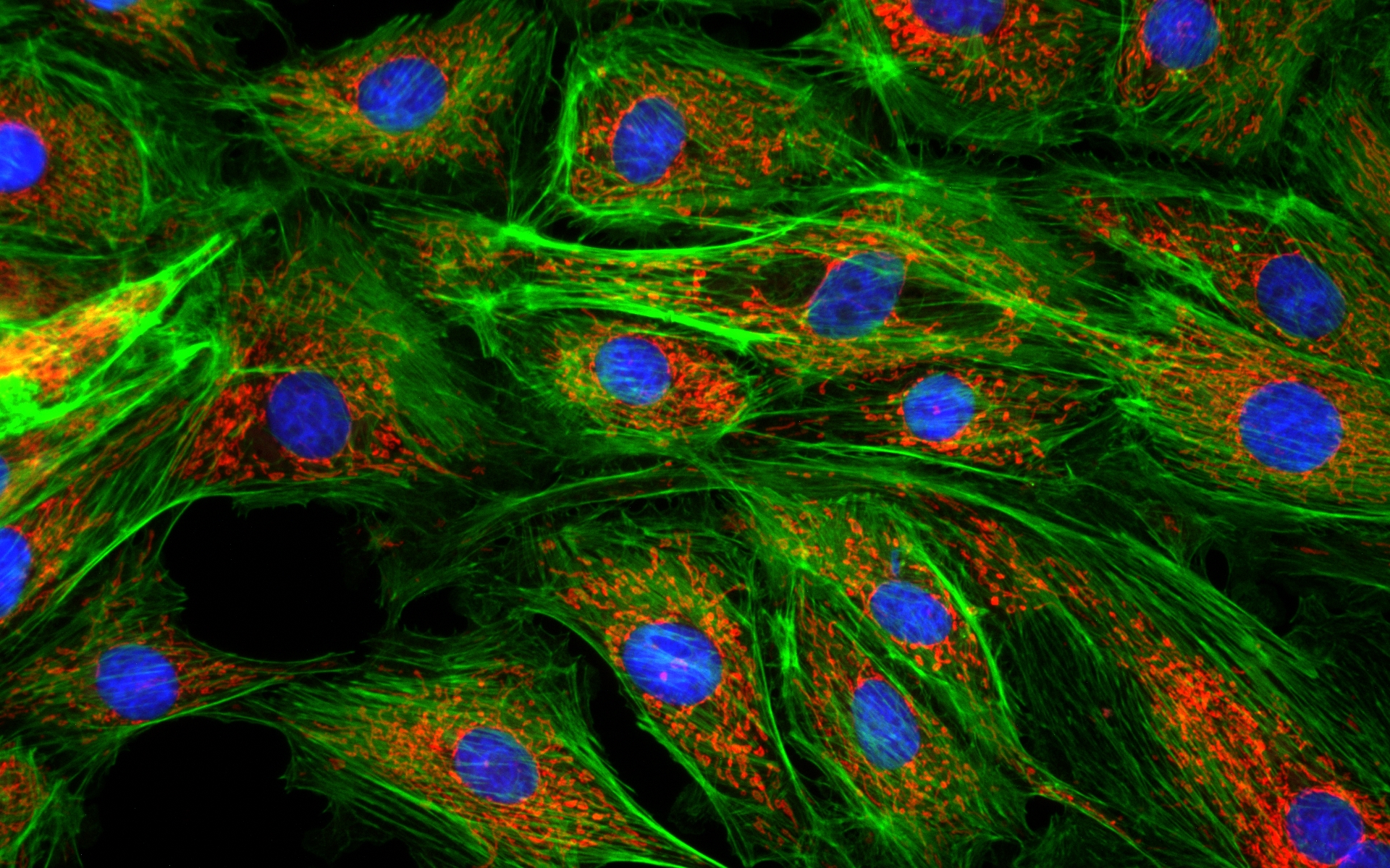
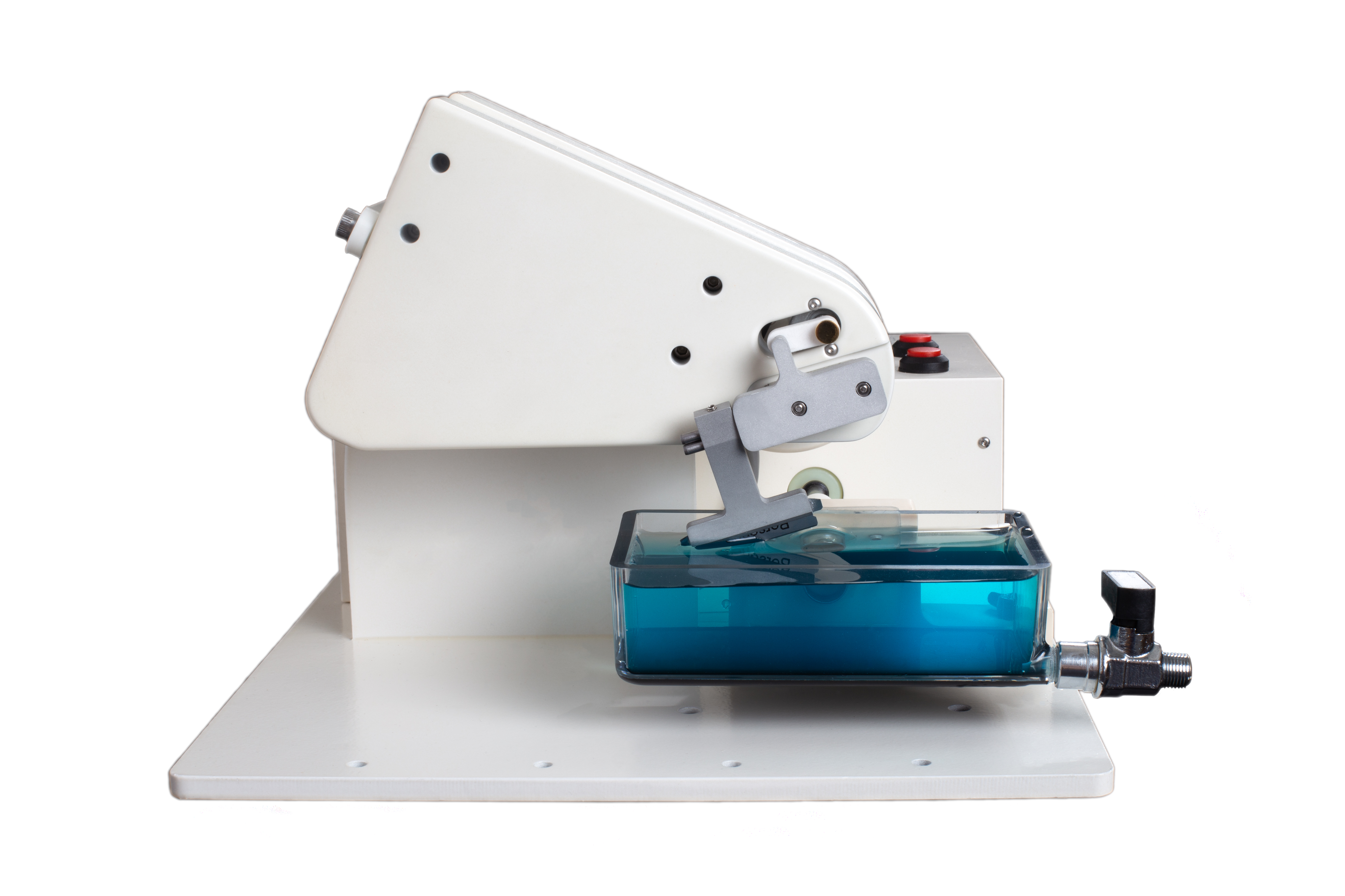
The pE-100 system has been developed for fluorescence applications requiring a single LED excitation wavelength.  It is perfect for clinical applications such as routine screening (e.g. using Auramine for Tuberculosis), for research applications requiring precise intensity control and fast-switching, or for electrophysiology applications where light has to be delivered to a specific location.  The user can select from 20 different LED wavelengths, ranging from the near-UV at 365nm to the near-IR at 770nm.  The system comprises a pE-100 LED Light Source, control pod, and power supply.

There are three standard pE-100 configuration options:
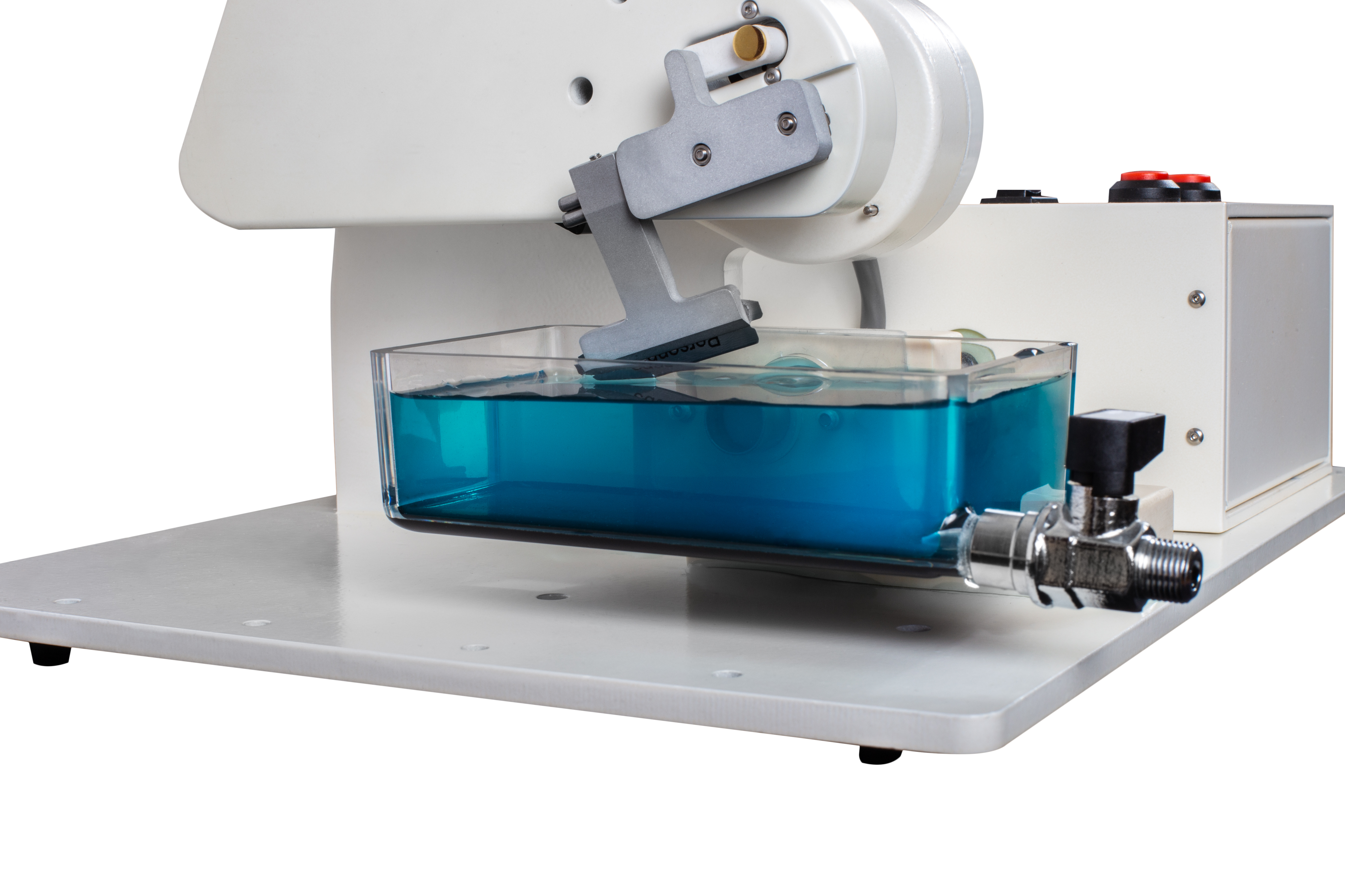
Direct-fit

(pE-100) for connecting to a microscope (epi-port) by selecting from a range of microscope adaptors  which covers all current and most older models.  A simple once only adjustment will allow optimisation to the optical path of the microscope.

Liquid Light Guide

(pE-100light guide) with a fixed 3mm diameter, liquid light guideFiber

(pE-100fiber) with an SMA connection for accepting multimode fibers.  The pE-100fiber has been designed with efficient coupling into a wide range of multimode fibers.  The pE-100fiber also includes excitation filter holders.

Combining pE-100s:

Direct fit configuration

pE-100 units can be combined using the pE-Combiner for applications requiring a second LED wavelength with independent control and triggering.

Liquid light guide or multimode fiberusing a fixed two wavelength configuration can be specified.  Two pE-100 Light Sources are combined, providing independent control and triggering. For further information go to the Downloads tab