analySIS LS
“analySIS“제품은 세계적으로 유명한 독일의 영상분석 전문업체인 OLYMPUS Soft Imaging Solution(OSIS) 사에서 제작한 최첨단 영상분석 프로그램입니다.
OLYMPUS Soft Imaging Solution사는 1987년 설립된 후 2006년 4월 OLYMPUS의 일원이 되었고 광학 현미경 및 전자 현미경용 분석 소프트웨어 및 디지탈 카메라 제품을 포함한 영상분석의 모든 솔루션 제공하고 있으며 유럽을 중심으로 다양한 분야에 사용되어 전세계의 많은 영상 분석 전문가와 연구진에게 사랑과 신뢰를 받고 있습니다.
주로 셀 카운팅, 크기 측정, 형광 관찰, 미생물 관찰 등과 같은 기능을 지닌 의학, 생명공학용 image analyzer인 analySIS LS는 다음과 같은 등급으로 구성되어 있고, analySIS LS Report 이상의 등급부터 필요한 module을 추가하거나 제품을 upgrade할 수 있습니다.
- LS STARTER : analySIS LS 의 첫 등급으로 형광 합성, 단순 측정과 같은 기능을 제공합니다.
- LS REPORT : analySIS LS의 두번째 등급으로서 STARTER의 모든 기능과 data base, report 기능이 추가 되었습니다. 또한 STARTER 이상의 등급부터 필요한 module을 추가하거나 제품을 upgrade할 수 있습니다.
- LS RESEARCH : analySIS LS의 세번쩨 등급으로서 REPORT의 모든 기능과 Microscope controller, Multiple Image Alignment (mia), multiple fluorescence imaging, Extended Focal Imaging (efi), Graph 등의 기능을 제공합니다.
- LS PROFESSIONAL : analySIS LS의 최고 등급으로서 RESEARCH의 모든 기능과 fft (fast fourier transformation), imaging C Track IT, Colocalisation 등의 기능을 제공합니다.
OLYMPUS Soft Imaging Solution 사의 최신 영상 분석 프로그램인 “analySIS LS” 시리즈는 사용자에게 정확하고 신뢰성 있는 데이터를 얻게 해 줄 것 입니다.
analysisFive LS Starter
analysisFive LS Starter는 entry 등급의 image analyzer로서 다음과 같은 기능을 수행할 수 있습니다.
Image Acquisition
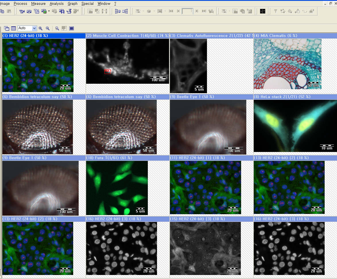
Olympus 및 OLympus Soft Imaging Solution의 모든 현미경용 카메라를 지원하며 완벽한 알고리즘을 통해 뛰어난 영상을 재현합니다. 이를 통해 획득한 영상은 최대 16장까지 동시에 비교 분석이 가능하면 동일 배율 지원, 동일 위치 지원과 같은 최상의 분석 기능을 제공합니다.
[동시비교-최대 16이미지]
Simple measurement
단순거리 측정, 수동 카운트, intensity 분석, 면적 분석 등과 같은 기능이 지원 됩니다.
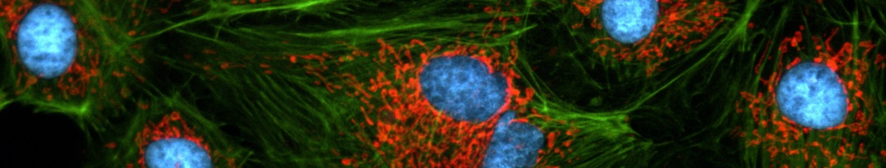
Multi-channel fluorescence
염색된 각각의 대상의 이미지를 한장씩 획득하여 하나의 이미지로 합성하여 쉽게 원하는 영상을 획득할 수 있습니다.
(olysia 홈페이지에서 동영상 파일 옮기기!)
analysisFive LS Report
analysisFive LS Report는 Starter의 모든 기능을 report작성, 이미지, 데이타의 데이타 베이스화 기능을 수행 할 수 있습니다.
Archiving(데이타 베이스)
새로운 STructured ARchive (STAR) database 는 이전의 Microsoft Access-style database를 대체합니다. 특히 Windows Explorer-type tree 구조에서 이미지 찾기를 보다 쉽게할 수 있도록 하였습니다. Database Assistant를 이용하여, database 구축을 쉽게할 수 있고 고급 기능을 통해 사용자의 취향에 맞는 정리가 가능합니다. 드래그와 드롭만으로 이 모든 기능이 자동으로 이루어 집니다. 그리고 모든 이미지는 사전에 정해놓은 폴더에 자동으로 저장할 수 있습니다.
(olysia 홈페이지에서 이미지 파일 옮기기!)
STAR databases can be queried in a variety of ways:
| • | simple queries of one or more fields with logical Boolean strings and wildcards |
| • | free queries |
| • | SQL queries |
데이타 베이스는 네트워크를 통해 자동 저장, 백업이 가능합니다.
(olysia 홈페이지에서 이미지 파일 옮기기!)
analysisFive LS Research
analysisFive LS Research는 Report의 모든 기능과 motorized 현미경 제어, new measurement, MIA, EFI 의 기능을 수행합니다.
Microscope & Hardware control
Olympus 현미경 BX61,IX81,SZX12/SZX16를 프로그램에서 제어할 수 있습니다.
이를 위해서는 하드웨어 마법사를 실행하여 motorized 현미경 장치-대물렌즈, 필터, 큐브(형광, dic), 콘덴서-를 구성하고 이는 프로그램 내부의 컴퍼넌트 리스트에서 쉽게 선택할 수 있습니다. 또한 여러 사용자마다 원하는 구성을 셋업도 할 수 있고 한번 셋업된 후에는 프로그램 작동만으로 모든 하드웨어들도 함께 자동으로 제어할 수 있습니다. 이를 통해 형광, 위상차, 편광, DIC 같은 여러 관찰도 한번의 셋팅과 클릭으로 편하게 사용할 수 있습니다.
(olysia 홈페이지에서 이미지 파일 옮기기!)
현미경 대물렌즈를 바꾸면 프로그램은 렌즈의 배율을 자동적으로 읽은 후 알맞은 스케일바를 이미지에 보여줍니다. 그리고 Z축이 motorized 되어 있으면 렌즈 변환시에도 촛점 거리를 자동으로 맞출 수 있습니다.
다음 회사 제품의 motorized stage를 지원합니다. Prior, Ludl, Märzhäuser
다음 회사 제품의 third-party shutters 와 filter wheels을 지원합니다.Prior, Ludl, Sutter, Uniblitz
(olysia 홈페이지에서 이미지 파일 옮기기!)
New measurement
새로운 측정툴은 매직완드, 다각형 측정, 자유 측정과 같은 강력한 기능을 지원하며 측정하고자 하는 값은 113가지가 가능합니다. (길이, 넓이, 반지름, intensity 등) 또한 측정된 값들은 측정 개체별로 통계값을 바로 볼 수 있으며, 아주 간단하게 sheet로 만들어 Microsoft Excel 형식 사용할 수 있습니다.
![]()
Dual Screen System
두 개의 모니터를 사용하여 넓은 화면으로 이미지의 분석 효율이 높아집니다.
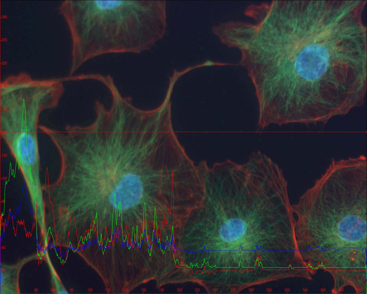
Graph
그래프 기능으로 레포트에 충실한 자료로 당신의 레포트 질을 높일 수 있습니다.
fis (Fast Image Acquisition)
고속으로 이미지를 획득하여 빠른 변화를 손쉽게 저장할 수 있습니다.
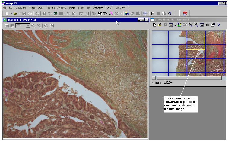
Stage Navigator
Stage Navigator를 이용하여 원하고자 하는 포인트를 지정하여 빠르고, 정확하게 확인 하실 수 있습니다.
With the Stage Navigator you can acquire overview images of a sample and use them for a precise navigation. This assures that you always know your exact location on the sample. You can recognize interesting areas in the sample, move to them in the live-image, and acquire images with a higher magnification. In the overview image, a “grid within a grid,” enables you to recognize the areas you have acquired with a higher magnification.
You can deactivate this display if you want to.
MIA (multiple image alignment)
관 여러장의 사진을 합성하여 넓은 영역을 하나의 파일로 만들어 편리한 관찰이 가능합니다.
Mia works with both monochrome and color images, supports the entire number of file formats in analySIS®, and includes an intelligent image acquisition mode with automatic calibration of the camera and camera and/or image rotation.
Once this has been defined, all you need to do is to make a decision about the required image size and resolution. Stage movement, image acquisition and the computation of the optimum overlap are done automatically by the software.
To effect the image montage, the individual images have to be aligned with sub-pixel accuracy. mia achieves this through intelligent pattern recognition techniques and plausibility checks within the overlap areas.
The resulting image has the same resolution as the individual images but is larger. It has more lines and columns. It thus represents an image that could not be acquired without mia with high resolution and large field of view.
How does Mia work?
1. The integrated image acquisition control allows the automatic acquisition of individual images; image rotation and stage displacement are automatically determined.
2. This example uses six individual images for the Mia process. There are no limits to the number of images Mia can process in one session. The number of images you actually use is dependent only on the storage capacity of your computer
3. After image acquisition, you can manually determine the number, pattern and correlation of the individual images. In the case of automatic image acquisition, these parameters will be preset for you.
4. After Mia has processed the images, the overlap areas can be adjusted automatically for differences in intensity.
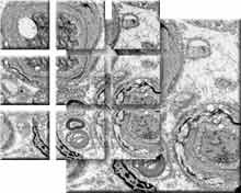
The result of the Mia process:
Image size:
2159 x 1062 pixels in 24-bit true color.
Nine image sections, each 1024 x 1024 pixels in size, automatically stiched together by the module Mia.
The high resolution 3300 x 3300 pixel sized full view image shows healthy and degenerated nerve cells.
EFI (extended focal imaging)
Focal depth 가 다른 이미지를 하나의 이미지로 합성하여 또렷한 이미지를 얻을 수 있습니다.
Microscopy at unlimited depth of focus
By using the efi module, you can solve a known and limiting problem of light microscopy: Microscopes in general have only a very limited depth of focus. Details which are visible in separate images with different focus settings are normally not visible in one single image.
efi records images with different focus settings and extracts those parts of the image that are in focus. Mounted into a single image, these details combine to create an image with unlimited depth of focus.
efi supports the automatic montaging of both color and b/w image series. Optionally, you can reconstruct a height map from these images. Used in live mode, efi provides both a live image as well as the partially reconstructed image. Missing details can be focused interactively and added to the current image.
efi automatically aligns images that show a lateral displacement, such as if the images were taken with a stereo microscope. Anoptional motor stage control provides the added advantage of automatic image acquisition and efi processing.
Real-time calculated recombination of image
efi circumvents the physical limitations of a microscope regarding its depth of focus. A number of images are recorded, each one with a different focus setting. The parts of each image that are in focus are extracted and combined into a single, focused image.
efi allows the combination of images with lateral displacements caused by different focus settings. This is a common effect when using a stereo microscope to capture the images. Before calculating the efi image, an integrated pre-alignment step shifts the images so that they overlap perfectly. Only after this alignment has been achieved does efi calculate the final image.
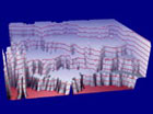
efi provides the option of calculating a height map from individual images. This height map can be used to produce a 3-D representation of the object. As an optional feature, the efi image can be used to texture the height map, creating a realistic topographic representation of the object’s surface.
A number of powerful functions for animating the 3-D structure within analySIS® enable the user to study the object from an optimal angle and position.
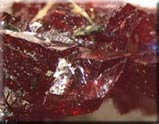
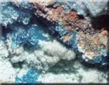
Real-time calculated recombination of image
Recombination of a focus series of a metallurgical sample. The recombined efi image is necessary for conducting advanced analysis.
efi allows the concurrent visualization of the live image directly from the microscope and the current recombined image. Areas that are still unfocused can be added interactively.
A simple addition of more images, even if the original acquisition was finished, allows adding further focused details to the image.
Real-time calculated recombination of image