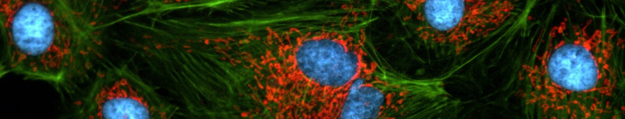
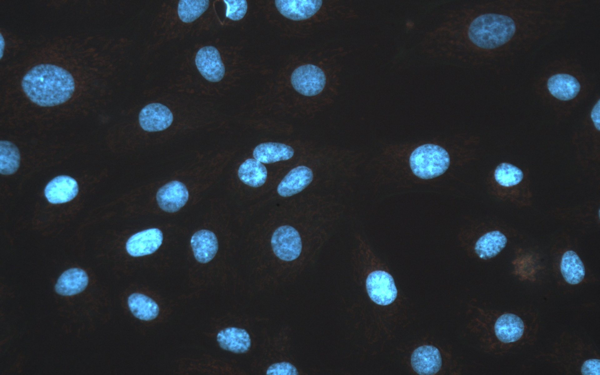
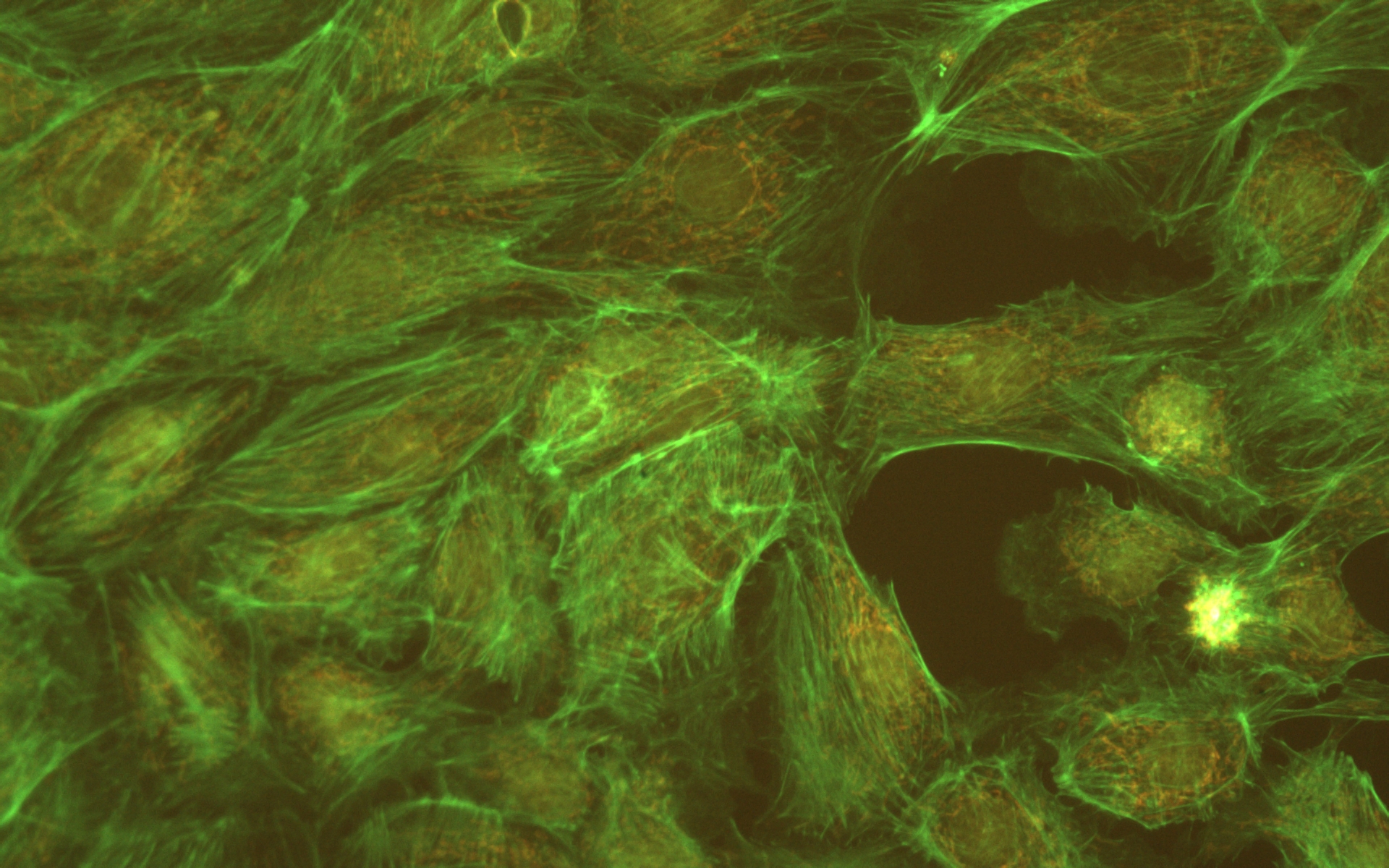
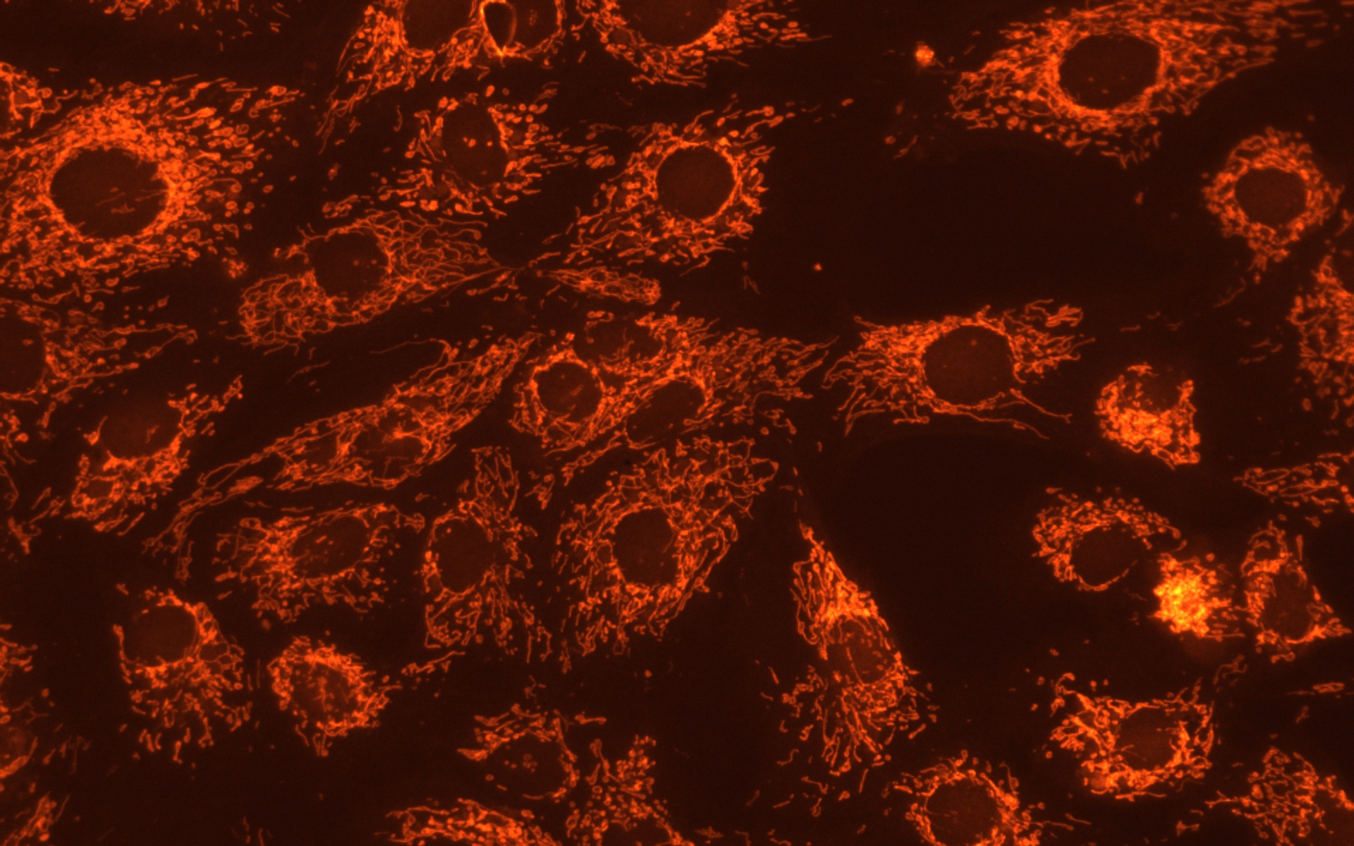
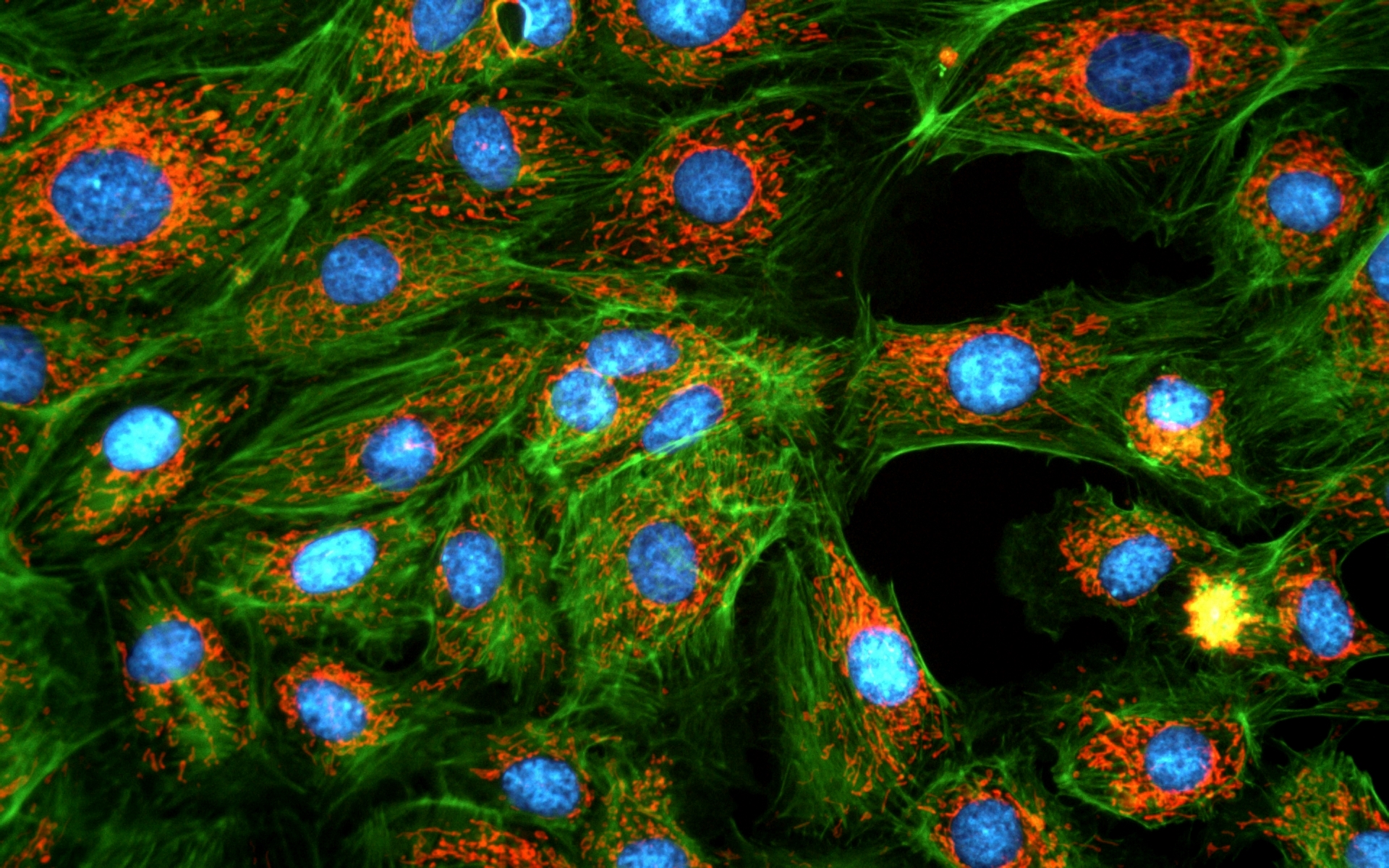
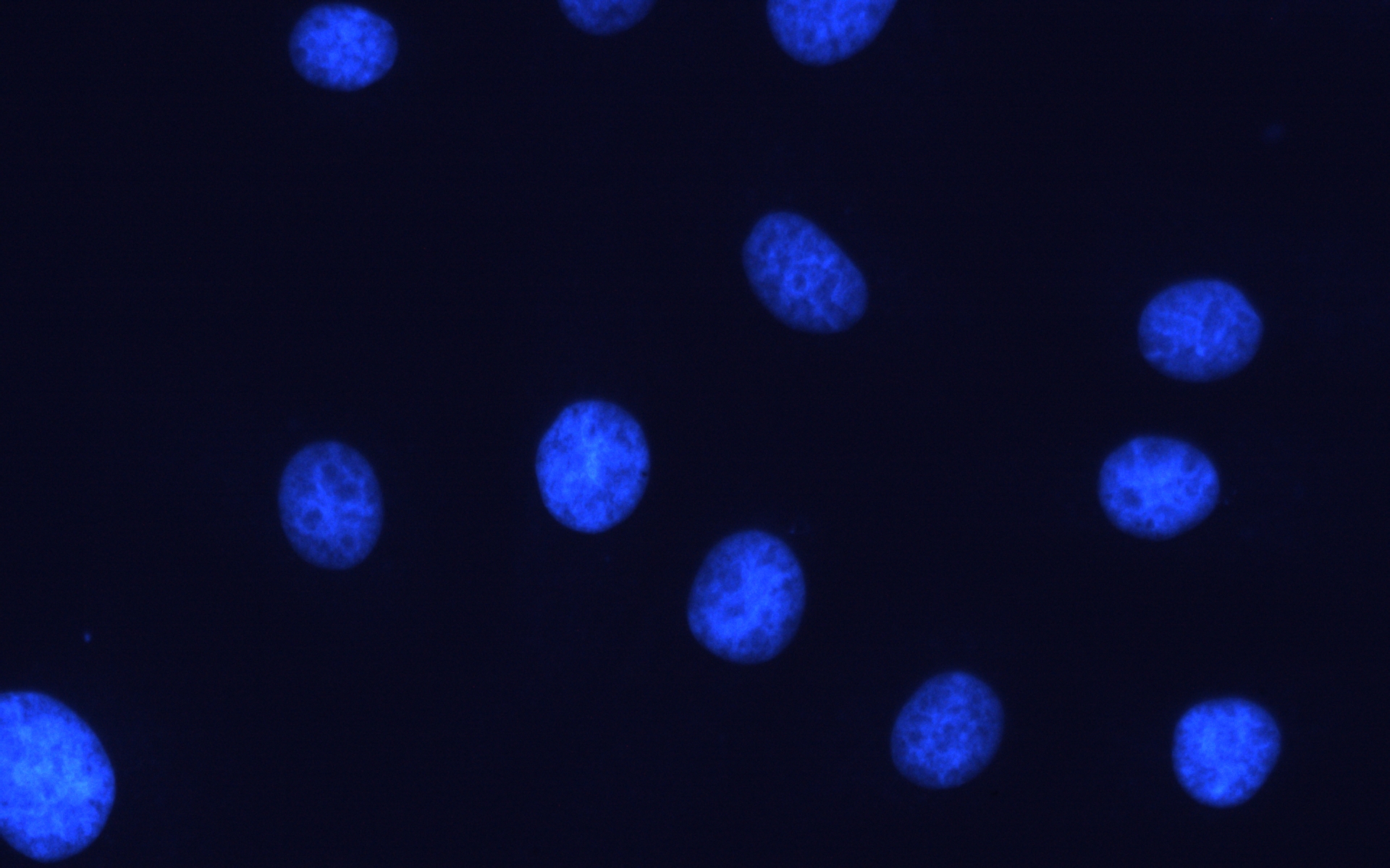
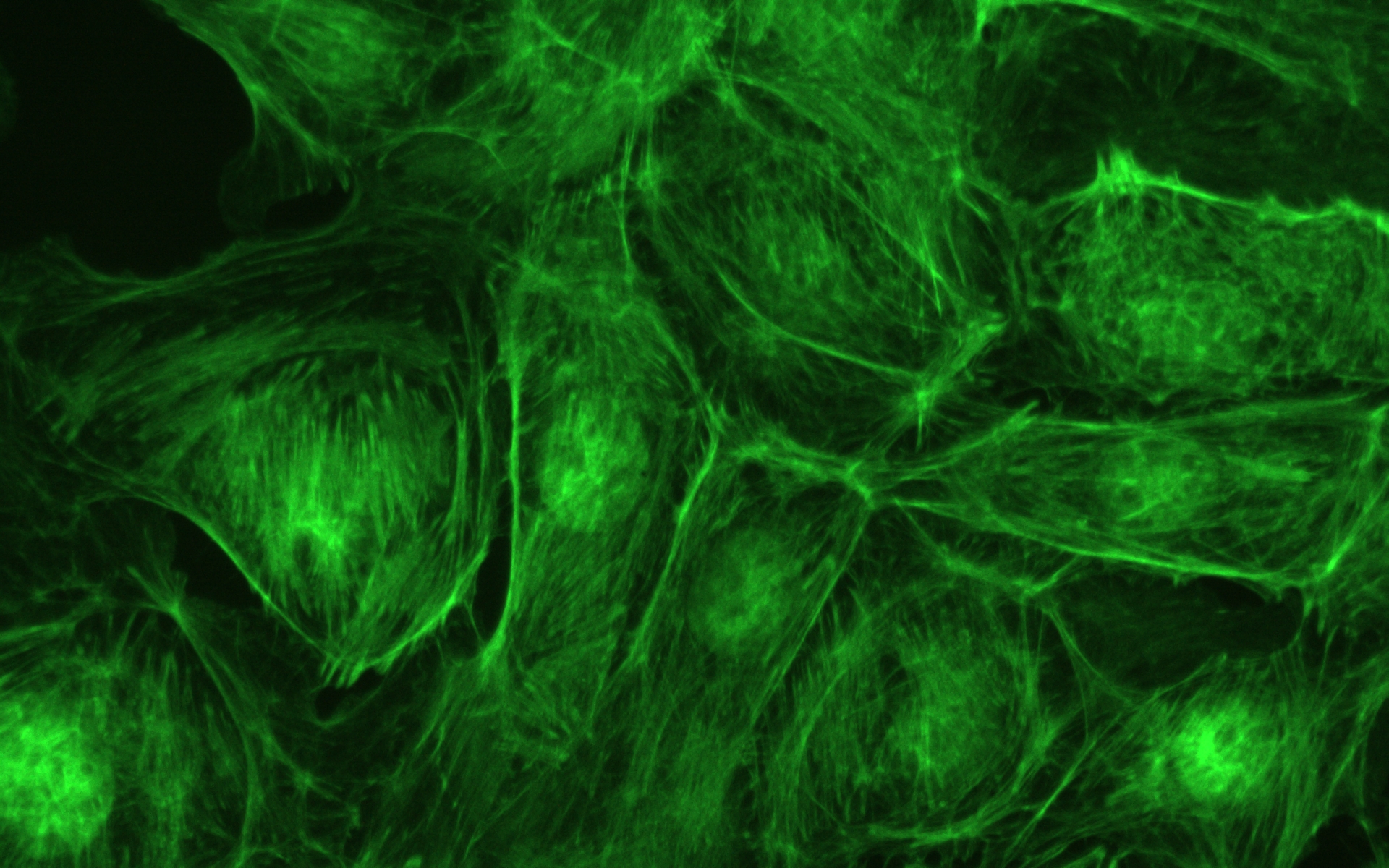
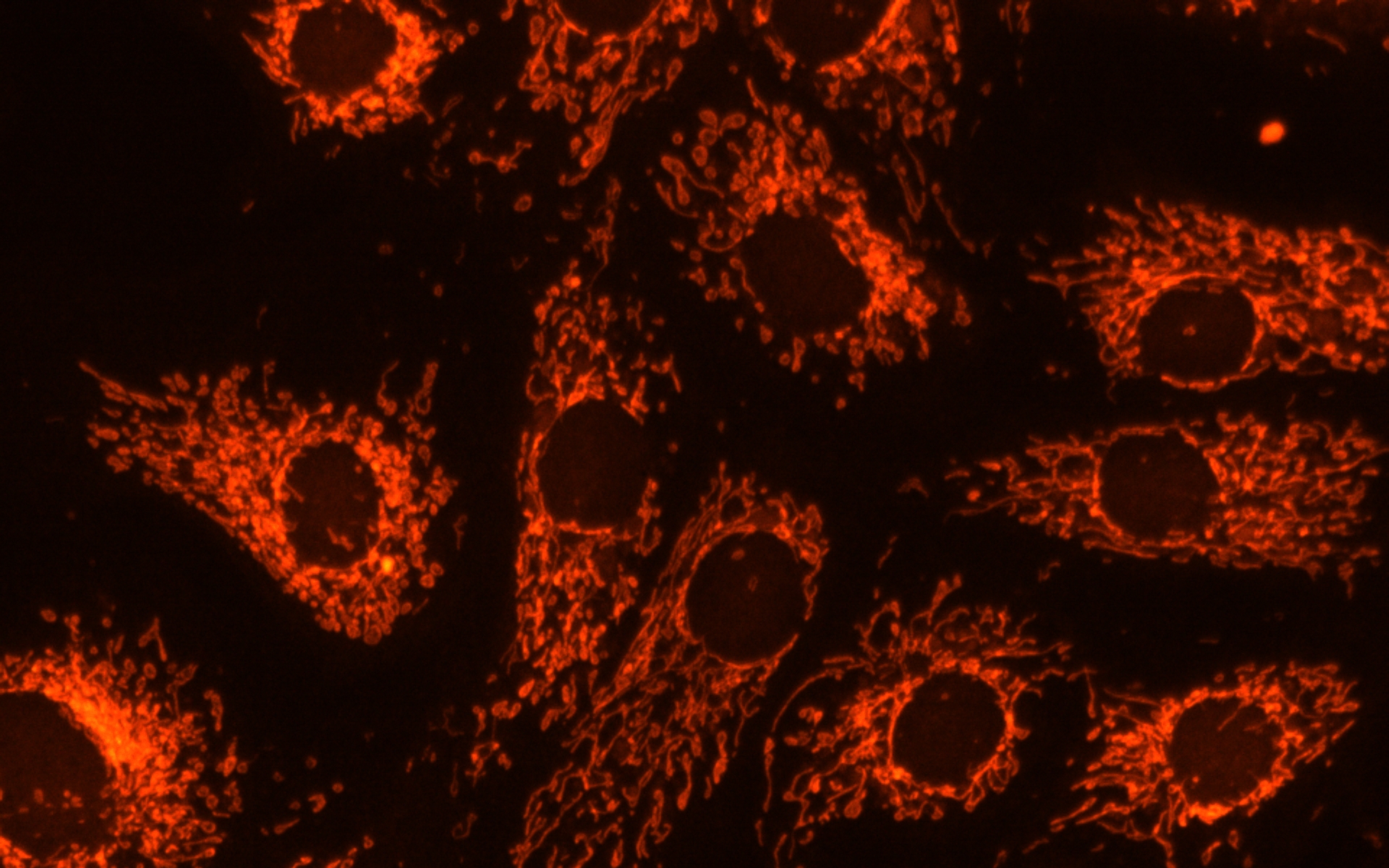
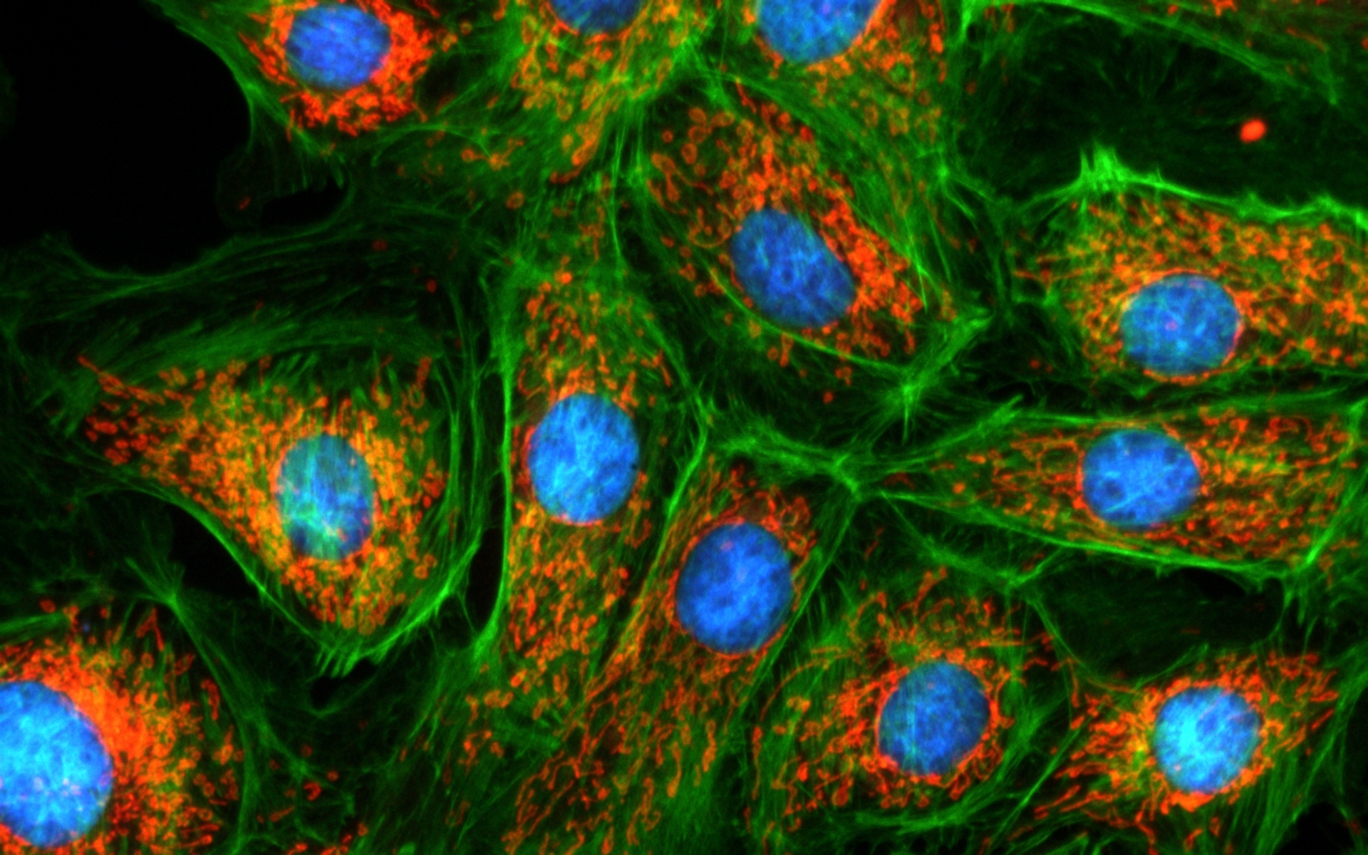
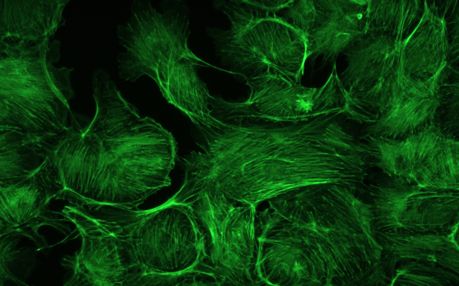
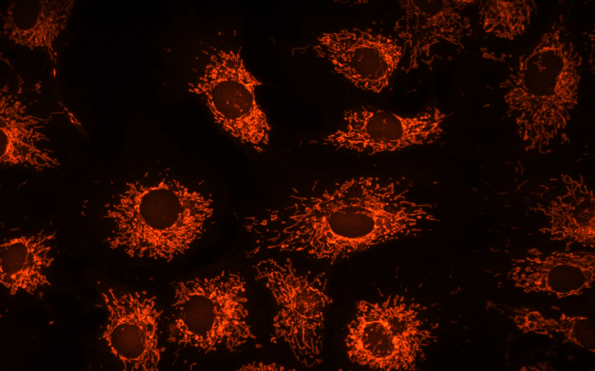
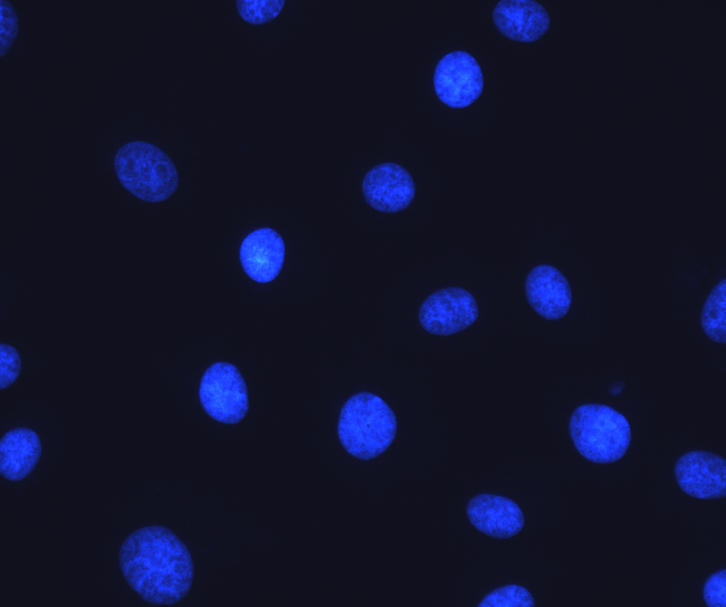
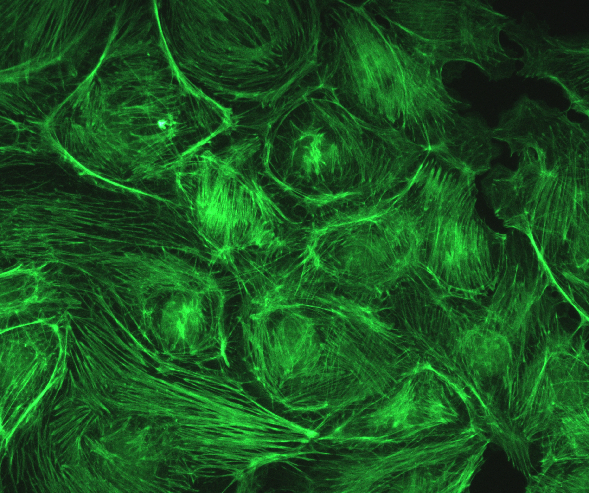
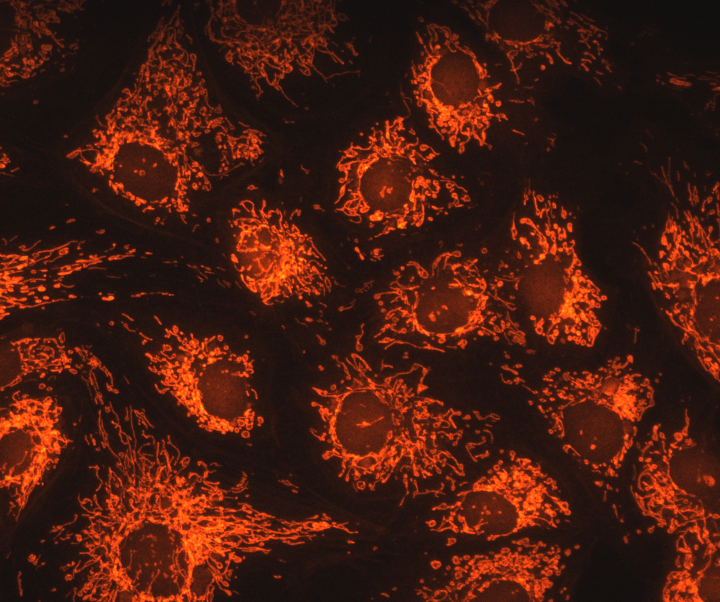
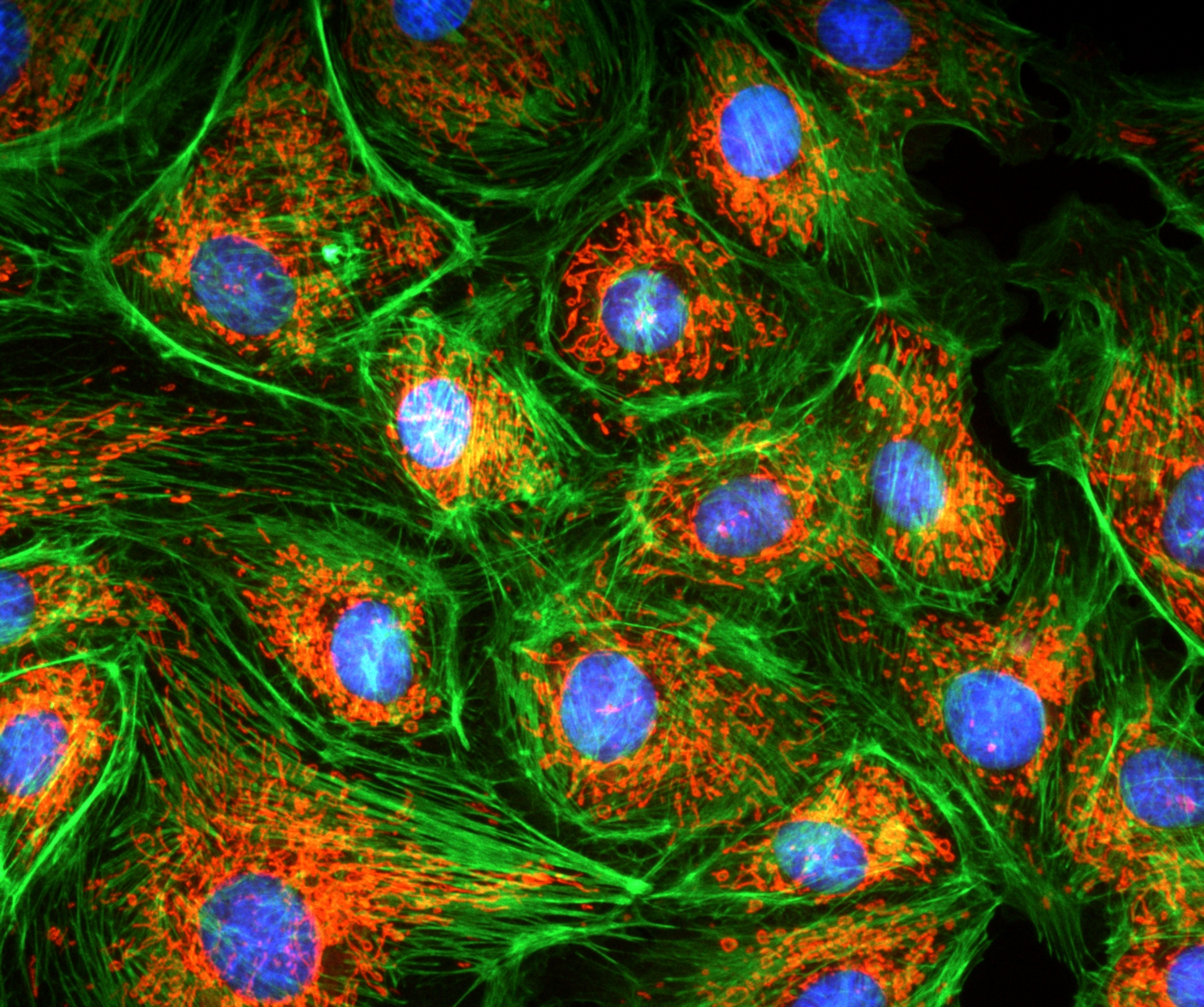
Sample : bovine pulmonary artery endothelial cell, BPAEC Filter Set : JNO-U(L), B(L), G(L) Camera : AcquCAM 23GR2 C-mount adapter : 1X Lens : UPLNAPO40X Above three images are merged and processed using ARM S/W
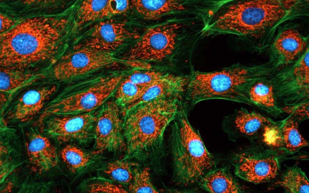
Sample : bovine pulmonary artery endothelial cell, BPAEC Filter Set : JNO-U(B), B(B), G(B) Camera : AcquCAM 23GR2 C-mount adapter : 1X Lens : PlanApo60xWLSM Above three images are merged and processed using ARM S/W
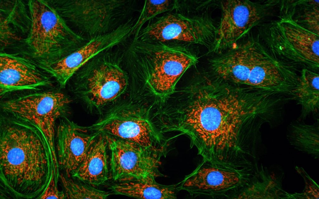
Sample : bovine pulmonary artery endothelial cell, BPAEC Camera : AcquCAM 23GR2 , Fluorescence Filter Set : JNO-U(B), B(B), G(B) Objective lens:UPLNAPO40X, C-mount adapter : 1X Above three images are merged and processed using JNO-ARM OLYMPUS BX51, UPlanApo40x U-TV1XC
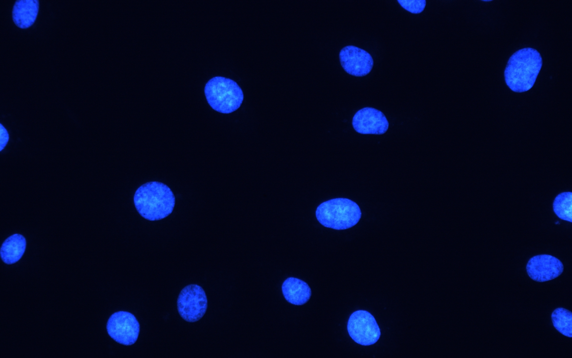
JNOpTIC AcquCAM 23GR2, JNO-UBG(B) C TYPE OLYMPUS BX51, UPlanApo40x U-TV1XC
JNOpTIC AcquCAM 23GR2, JNO-UBG(B) B TYPE
USB 3.0 Color Camera
1/1.2 ” Exmor Sensor
1920×1200 pixel
Trigger input and I/O
Available of taking images with a wavelength of 650nm or more, such as CY5
High Sensitivity Sensor (Best)
Available of images with a very wide field of view
The displays live digital images with gradual smoothness and combines exceptional resolution with faithful color reproduction.The new sensors feature a global shutter function and able to capture a high-speed moving image without focal plane distortion. High-speed processing, low noise and low power dissipation by using column-parallel A/D conversion. equipped with trigger mode, and the external pulse can control accumulation time. The Sensor also have a pulse output function to indicate respective conditions during shutter operation and can be coordinated with peripheral circuits. High-definition images can be displayed live at a rate of high frames per second. without compression. Such imaging quality enables even the finest cellular regions to be observed clearly and distinctly without deterioration. While focusing is made stress free.
Sample : bovine pulmonary artery endothelial cell, BPAEC Filter Set : JNO-U(B), B(B), G(B) Camera : AcquCAM 23GR2 C-mount adapter : 1X Lens : UPLNAPO40X Above three images are merged and processed using ARM S/W
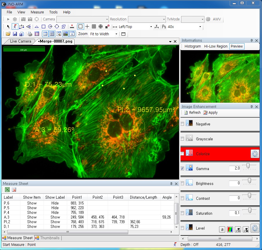
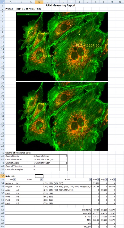
ARM is image analysis software for JNOPTIC AcquCAM cameras. This S/W is interchangeable with all WDM cameras regardless of camera brand and model and even more the most strength point is simple and easy use of length, area, angle, etc.
2. Simple measurement tools
Measurement Tools: Count, Distance, Angle, Area, etc.
Available measurement on LIVE image and saved image mode
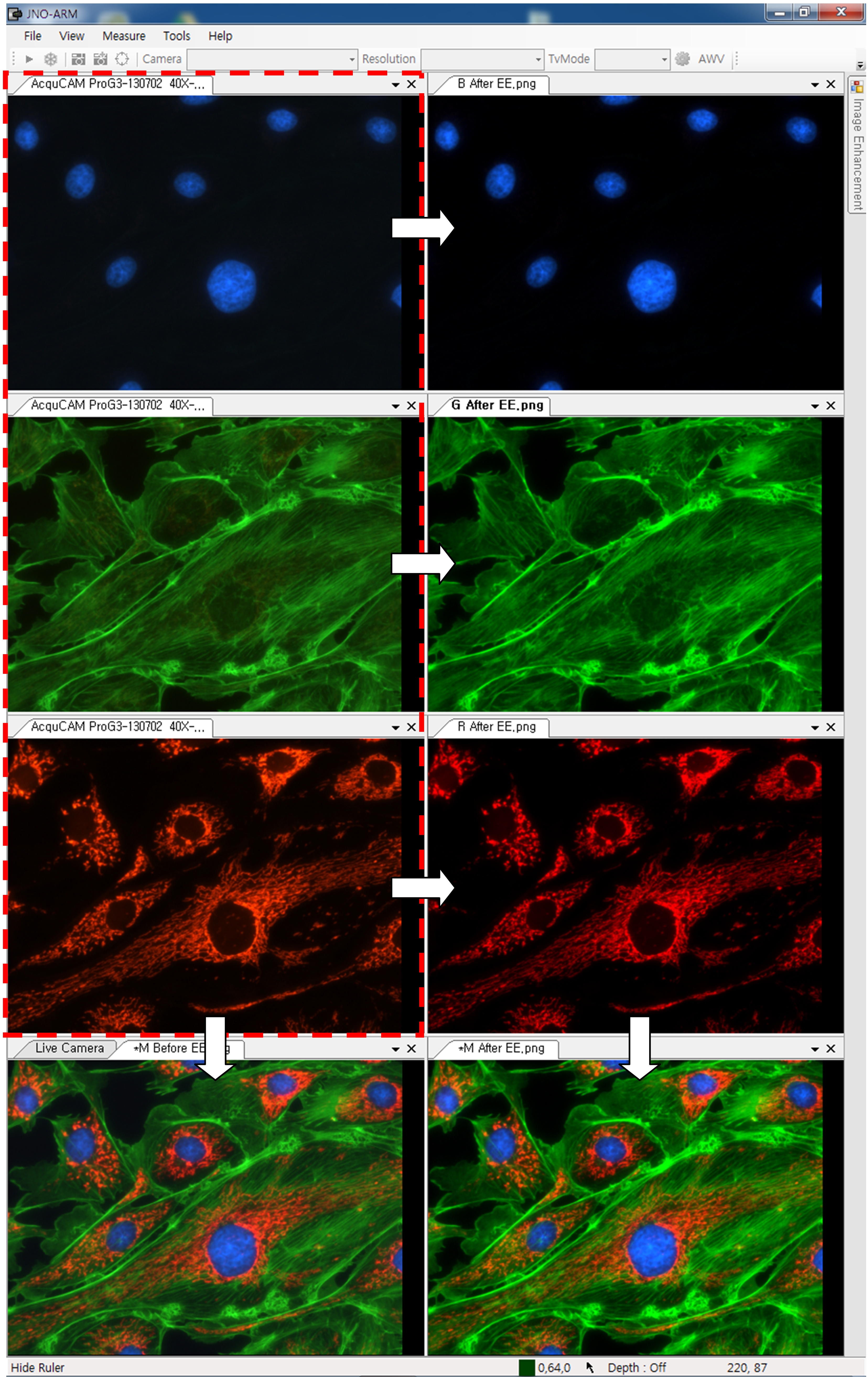
3-1. Specialization for observation of fluorescence images
Improvement Function about Fluorescence Microscopy Image. (Used function: Auto Level, Low level, Pseudo Color, Merge Image)
Bottom on the left is merged image without improvement of image. And the bottom on the right is merged image after improvement of image. These images are shot by JNOPTIC AcquCAM Pro/G3 camera
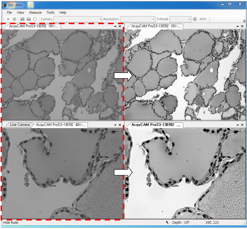
3-2-1 Effect of high-contrast image with simple operation (Monochrome)
Improvement Function about Microscopy Image Monochrome. (Used function: Auto Level)
Effective improvement of image with simple operation. This image is shot by JNOPTIC Pro/S3 camera
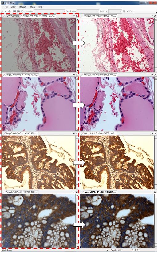
3-2-2 Effect of high-contrast image with simple operation (Color)
Improvement Function about Microscopy Image. (Used function: Auto Level)
Effective improvement of image with simple operation. These images are shot by JNOPTIC AcquCAM Pro/G3 camera
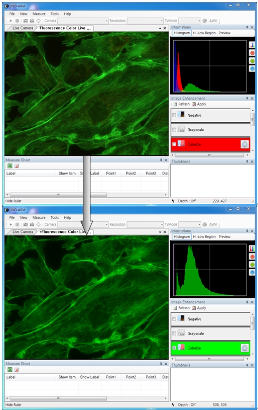
3-3-1 Live Pseudo Color (for color camera)
When you get fluorescence images using color camera, if the unwanted channel was shown because of cross talk, you could remove this channel to take advantage of function of Live Pseudo.
Improvement Function about Fluorescence Microscopy Live Camera Image. (Used function: Live Pseudo Color)
The extraction of wanted color information from color image (Remove BR on RGB source) These images are shot by JNOPTIC AcquCAM Pro/G3 camera
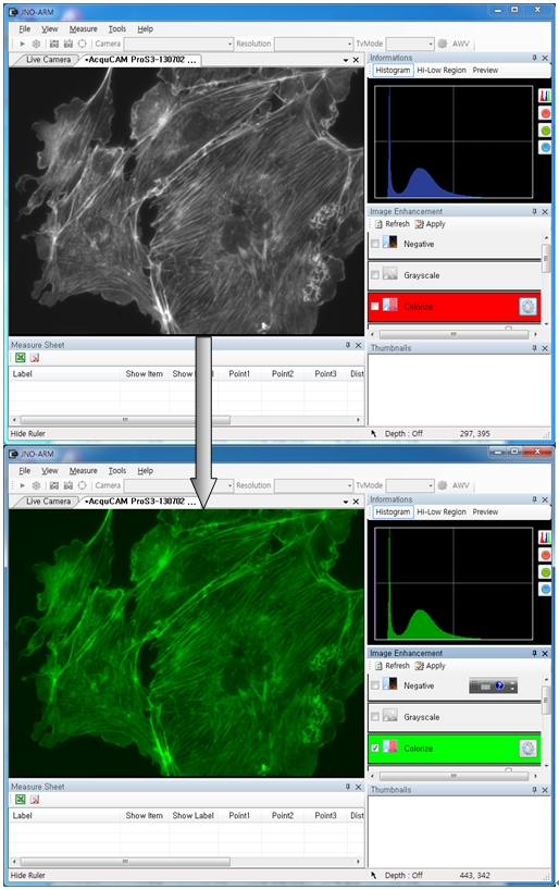
3-3-2 Live Pseudo Color (for Monochrome camera)
When you get fluorescence images using monochrome camera, you can raise efficiency of acquisition for fluorescence images to add similar false color to the color to be shown on the eyepiece in real time.
Improvement Function about Fluorescence Microscopy Live Monochrome Camera Image. (Used function: Live Pseudo Color)
Improvement of usage environment for monochrome camera to add false color on the image. These images are shot by JNOPTIC AcquCAM Pro/S3 camera
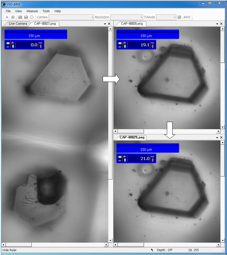
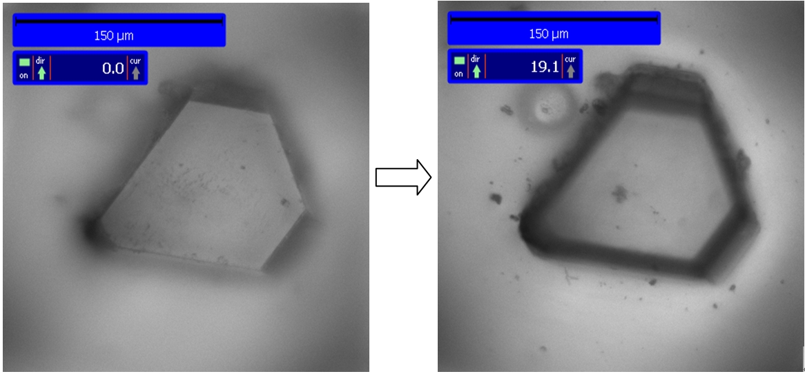
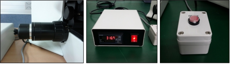
4. JNO-MHU(Option Unit)
Available measuring height with additional unit, JNO-MHU
※ JNO-MHU is sold separately as optional unit.
Left: Focus on top of sample (Z-axis reset), Right: Focus on bottom of sample (Z-axis height measurement)
< Measuring condition : over 20x objective, temp 20°C >
Available of images with a very wide field of view
AcquCAM 5G color camera has a USB 3.0 interface and is the perfect solution for many industrial automation, quality assurance, security, surveillance and medical applications. The color camera ships with the very sensitive 2/3 inch CMOS Pregius sensor. With up to 38 images per second, the AcuqCAM 5G is a low cost, yet highly versatile imaging solution. The camera includes a C to CS mount adapter, making it compatible to C and CS mount lenses. Using the optional CS to M12 board lens adapter, the camera is
also compatible to M12 board lenses.
Sample : bovine pulmonary artery endothelial cell, BPAEC Filter Set : JNO-U(B), B(B), G(B) Camera : AcquCAM 5G C-mount adapter : 1X Lens : UPLNAPO40X Above three images are merged and processed using ARM S/W
ARM is image analysis software for JNOPTIC AcquCAM cameras. This S/W is interchangeable with all WDM cameras regardless of camera brand and model and even more the most strength point is simple and easy use of length, area, angle, etc.
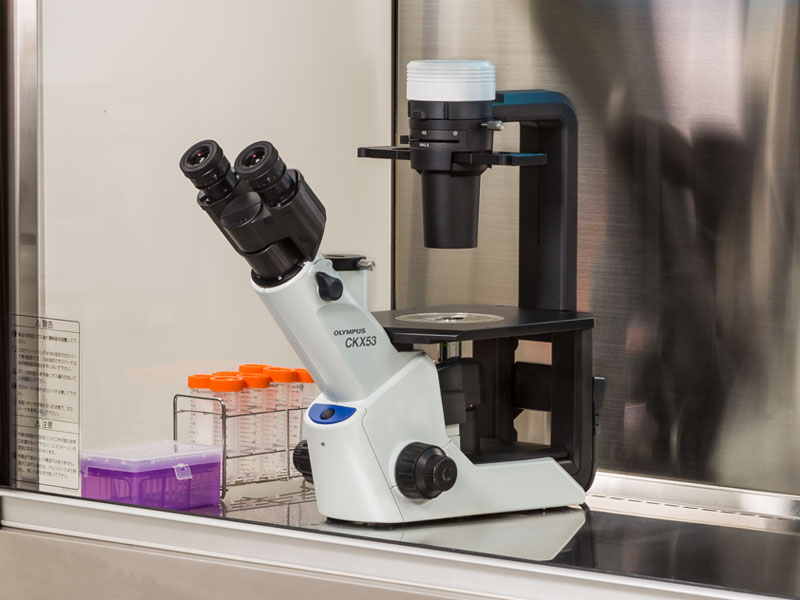
향상된 이미지 품질과 인체공학 설계로, Olympus CKX53은 라이브 셀 관찰, 세포 샘플링 및 처리, 이미지 캡처, 그리고 형광 관찰을 포함한 다양한 세포 배양 샘플에 뛰어난 성능과 효율적인 관찰 흐름을 제공합니다.
라이브 셀 관찰
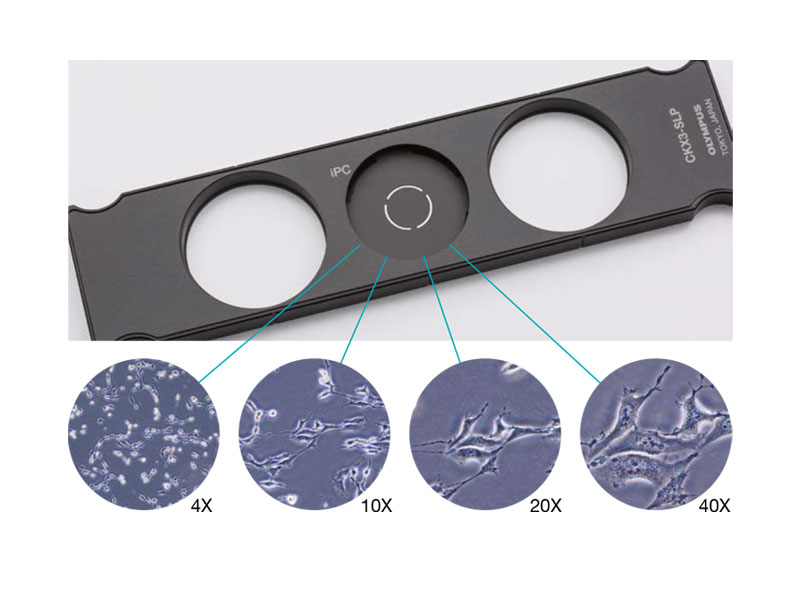
통합 위상차( iPC : Integrated Phase Contrast ) 현미경
CKX53 iPC 시스템을 이용하여 대물렌즈 배율( 4x, 10x, 20x , 40x ) 변경시, 콘덴서 측의 위상차 링슬릿의 연동 변경이 필요 없게 되어 효율적인 관찰 작업이 가능하고, 또한 위상차 설정이 틀어짐에 대한 수시 조정이 필요없어 언제나 선명한 샘플 관찰이 가능합니다.
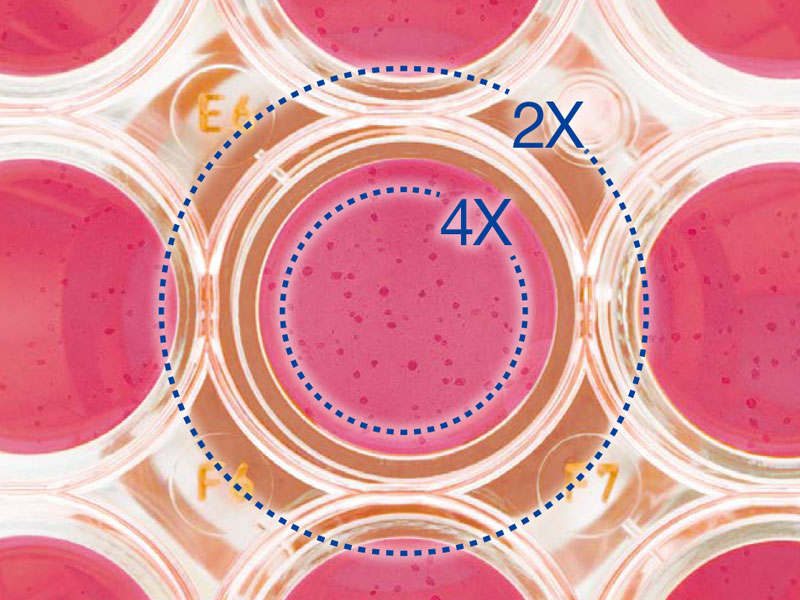
2X 배율, FN 22의 대물렌즈로 선명하고 넓은 시야
PLN2X 대물렌즈를 위한 링 슬릿, CKX3-SLPAS는 직경 11 ㎜ , 시야수 22 ㎜ 를 갖습니다.
2X 대물렌즈는 다른 대물렌즈 보다 확연히 높은 contrast를 제공하여, 투명한 샘플도 명확하게 식별할 수 있습니다. 예를 들어, 96-웰 마이크로 플레이트 관찰시, 넓은 시야로 인하여 스테이지를 움직이지 않고 웰의 모든 세포를 관찰 할 수 있습니다.
IVC (Inversion Contrast) 기술을 사용한 3D 셀 관찰
새로 개발된 IVC 기술로, 위상차보다 시야 심도는 좁아지며, 개체의 모양이나 투명도와 관계없이 삼차원 이미지를 선명하게 합니다. 또한, IVC 관찰은 후광 효과나, 방향성 있는 그림자를 배제하여, 개체의 선명한 관찰을 가능하게 합니다. * 10X 대물렌즈 (PLCN10X, CACHN10XIPC)는 새로운 IVC 관찰에 사용할 수 있습니다.
Glass Heater for microscope
TPi-CKX53X ( Thermo Glass Plate )
Microscope:Olympus CKX53 series
Applicable stage: XY mechanical stage CKX3-MVR
Setting range: ambient ~ 60℃
Plate dimension: W190 x D138㎜
Heating area: W174 × D127㎜
Glass thickness: 0.5 ㎜
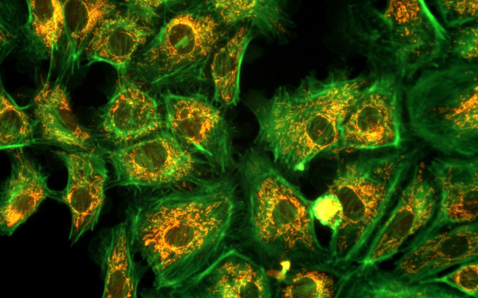
형광 관찰 (Fluorescence Microscopy)
다양한 형광 시약과 선명한 시야
100 W 수은 램프 (U-LH100HG), 130 W 고압 수은 램프 (U-HGLGPS), 그리고 타사(3rd Party) LEDs*와 같은 여러 통합 광원을 이용하여 형광 이미지를 선명하게 관찰할 수 있습니다. 일반 연구용 형광현미경 IX3 및 BX3 에서 사용하는 미러 유닛을 동일하게 사용 할 수 있습니다.
3개의 형광미러 유닛을 장착할 수 있으나, Bright Field 관찰과 위상차 관찰에 영향을 줄 수 있으니 유닛 선택시 고려할 필요가 있습니다.
Image taken by AcquCAM 23GR2 with LUCPlanFLN40x Ph2, 1x Adapter, CKX53
밝은 조건에서 높은 Contrast
“Umbra Shield”는 특히 CKX53을 사용한 형광 관찰을 위해 설계되었습니다. 차단막은 실내 광원을 효과적으로 차단하여 형광의 대비를 향상하여 밝은 실험실 조건에서도 선명한 형광 관찰이 가능합니다. 위상차를 사용하는 경우, Umbra 차단막을 들어 올려 표본에 빛을 통과시킬 수 있습니다.
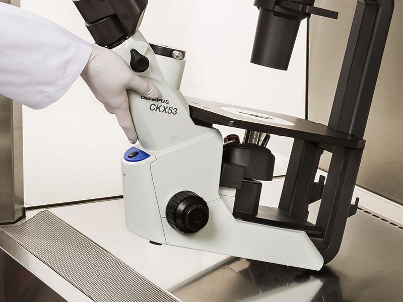
CKX53 현미경은 UV 차단 코팅 덕분에 UV 살균 공정 중에 그대로 둘 수 있습니다. 이 시스템은 약 7kg (15.4lb)으로 이전 모델보다 가볍고 설치 공간이 더 작기 때문에 실험실 공간을 덜 차지합니다. 또한, 한 손으로 현미경을 움직일 수 있으며 관찰 경통의 목 부분을 이용하여 쉽게 운반 할 수 있습니다.
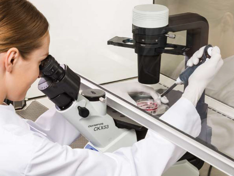
멸균 벤치 환경에서 간편한 세포 샘플링
CKX53의 접안렌즈와 광축/포커스 노브 사이의 거리가 짧으므로 작업자의 손의 위치를 자연스럽게 잡을 수 있어서 초점 및 셀 샘플링이 용이합니다.
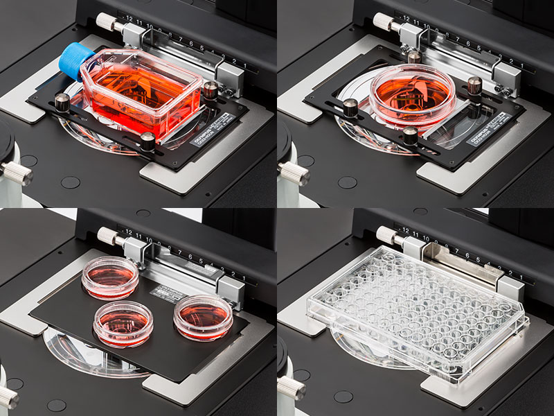
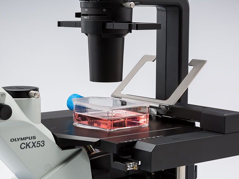
다양한 세포 배양 용기를 사용할 수 있습니다.
CKX53의 공용 홀더로, 디쉬, 마이크로플레이트, 플라스크를 포함한 다양한 용기에서 배양된 세포를 확인하기 쉽습니다. 옵션 홀더가 부착되면, 최대 세 개의 35㎜ 배양 용기를 스테이지에 장착할 수 있습니다. 또한, 다양한 마이크로플레이트를 별도의 홀더 없이 다룰 수 있습니다.
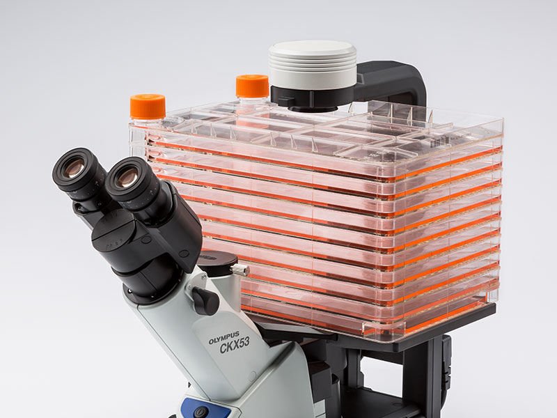
다층 조직 플라스크(Multi-Layer Tissue Flask)를 위한 종합적인 관찰
CKX53의 폭과 탈착 가능한 콘덴서로 다층 조직 플라스크와 같은, 최대 190 ㎜ 높이의 배양 용기도 볼 수 있습니다. PLCN4X 대물렌즈의 우수한 초점 심도로 다층 조직 플라스크 내 바닥 두 개의 층의 세포를 빠르고 편하게 관찰할 수 있습니다.
다양한 용기를 사용하여 관찰 유연성 증대
홀더 암을 들어 올려서 수동으로 세포 배양 용기를 배치할 수 있습니다. 또한, 스테이지는 좌우로 최대 70 ㎜ 까지 확장할 수 있습니다.